Introduction to Bioethics: The Boundary between Research and Clinical Practice
Special Report | Volume 1 | Issue 1 | JRS December 2021 | Page 48-50 | Graciela Moya. DOI: 10.13107/jrs.2021.v01.i01.025
Author: Graciela Moya [1]
[1] Department of Bioethics, Facultad de Ciencias Médicas, Pontificia Universidad Católica Argentina, Argentina, South America.
Address of Correspondence
Dr. Graciela Moya, MD, PhD.
Instituto de Bioética, Pontificia Universidad Católica Argentina, Argentina, South America.
E-mail: gracielamoya@uca.edu.ar
References:
1. Gelijns AC, Rosenberg N, Moskowitz AJ. Capturing the unexpected benefits of medical research. N Engl J Med 1998;339:693-8.
2. McCormick JB, Sharp RR, Ottenberg AL, Reider CR, Taylor HA, Wilfond BS. The establishment of research ethics consultation services (RECS): An emerging research resource. Clin Transl Sci 2013;6:40-4.
3. Sharp RR, Taylor HA, Brinich MA, Boyle MM, Cho M, Coors M, et al. Research ethics consultation: Ethical and professional practice challenges and recommendations. Acad Med 2015;90:615-20.
4. Grodin MA, Annas GJ. Legacies of Nuremberg: Medical ethics and human rights. JAMA 1996;276:1682-3.
5. Annas GJ, Grodin MA, editors. The Nazi Doctors and the Nuremberg Code: Human Rights in Human Experimentation. New York: Oxford University Press; 1992.
6. U.N. Universal Declaration of Human Rights. Available from: https://www.un.org/en/about-us/universal-declaration-of-human-rights [Last accessed on 2021 Oct 13].
7. Rice TW. The historical, ethical, and legal background of human-subjects research. Respir Care 2008;53:1325-9.
8. World Medical Association, Declaration of Helsinki, Ethical Principles for Medical Research Involving Human Subjects. Available from: https://www.wma.net/policies-post/wma-declaration-of-helsinki-ethical-principles-for-medical-research-involving-human-subjects [Last accessed on 2021 Oct 13].
9. World Health Organization. Research Ethics Committees: Basic Concepts for Capacity-building. Geneva: World Health Organization; 2009. Available from: https://www.who.int/ethics/ethics_basic_concepts_eng.pdf [Last accessed on 2021 Oct 13].
10. Council of Europe, Steering Committee on Bioethics. Guide for Research Ethics Committee Members; 2010. Available from: https://www.coe.int/t/dg3/healthbioethic/activities/02_biomedical_research_en/Guide/Guide_EN.pdf [Last accessed on 2021 Oct 13].
11. The National Commission for the Protection of Human Subjects of Biomedical and Behavioral Research. Belmont Report, Ethical Principles and Guidelines for the Protection of Human Subjects of Research. Available from: https://www.hhs.gov/ohrp/regulations-and-policy/belmont-report/read-the-belmont-report/index.html [Last accessed on 2021 Oct 13].
12. CIOMS International Ethical Guidelines for Health-related Research Involving Humans. Geneva: CIOMS; 2016. Available from: https://www.cioms.ch/wp-content/uploads/2017/01/WEB-CIOMS-EthicalGuidelines.pdf [Last accessed on 2021 Oct 13].
13. CIOMS Clinical Research in Resource-limited Settings. Geneva: CIOMS; 2021. Available from: https://wwww.cioms.ch/publications/product/clinical-research-in-low-resource-settings [Last accessed on 2021 Oct 13].
14. International Council for Harmonisation of Technical Requirements for Pharmaceuticals for Human Use. Available from: https://www.ich.org [Last accessed on 2021 Oct 13].
15. Emanuel EJ, Wendler D, Grady C. What makes clinical research ethical? JAMA 2000;283:2701-11.
16. Sugarman J. Methods in Medical Ethics. Washington, DC: Georgetown University Press; 2001.
17. Martinson B, Anderson M, de Vries R. Scientists behaving badly. J Nat 2005;435:737-8.
18. Amer A. The health care ethics: Overview of the basics. Open J Nurs 2019;9:183-7.
19. Samuel G, Chubb J, Derrick G. Boundaries between research ethics and ethical research use in artificial intelligence health research. J Empir Res Hum Res Ethics 2021;16:325-37.

| How to Cite this article: Moya G | Introduction to Bioethics: The Boundary between Research and Clinical Practice | Journal of Regenerative Science | Dec 2021; 1(1): 48-50. |
[Full Text HTML] [Full Text PDF] [XML]
Multilineage-differentiating Stress-enduring (MUSE) Cells in Orthobiologics: Are they the Future?
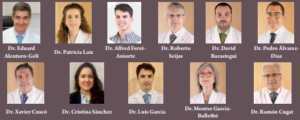
Review Article | Volume 1 | Issue 1 | JRS December 2021 | Page 44-47 | Eduard Alentorn-Geli, Patricia Laiz, Alfred Ferré-Aniorte, Roberto Seijas, David
Barastegui, Pedro Álvarez-Díaz, Xavier Cuscó, Cristina Sánchez, Luís García, Montse García-Balletbó, Ramón Cugat. DOI: 10.13107/jrs.2021.v01.i01.023
Author: Eduard Alentorn-Geli [1,2,3], Patricia Laiz [1,2], Alfred Ferré-Aniorte [1,2], Roberto Seijas [1,2], David Barastegui [1,2,3], Pedro Álvarez-Díaz [1,2,3], Xavier Cuscó [1,2], Cristina Sánchez [1,2], Luís García [1,2], Montse García-Balletbó [1,2], Ramón Cugat [1,2,3]
[1] Instituto Cugat, Hospital Quironsalud. Plaza Alfonso Comín 5-7, Planta -1, 08027 Barcelona, Spain.
[2] Fundación García Cugat, Plaza Alfonso Comín 5-7, Planta -1, 08027 Barcelona, Spain. Barcelona, Spain.
[3] Mutualidad de Futbolistas (Real Federación Española de Fútbol), Delegación catalana. Ronda Sant Pere 19-21, Entresuelo, 08010, Barcelona, Spain.
Address of Correspondence
Dr. Ramón Cugat Bertomeu, MD, PhD,
Instituto Cugat, Plaza Alfonso Comín 5-7, 08023 Barcelona, Spain.
E-mail: ramon.cugat@sportrauma.com
Abstract
Multilineage-differentiating stress-enduring (MUSE) cells are non-tumorigenic pluripotent stem cells with endogenous reparative properties. These cells have a very powerful ability to adapt to global environment changes and are thus stress-tolerant cells. Interestingly, MUSE cells can differentiate into cells representative of all three germ layers. There has been a number of studies demonstrating its powerful regenerative power in several disorders: type-1 diabetes mellitus, myocardial infarction, stroke, glomerular-related kidney diseases, chronic liver failure, and ischemia-reperfusion lung injury. Recent data have also suggested that MUSE cells have significant repair properties for osteochondral lesions. The present article will review what are MUSE cells and how they work, the application of these cells into different disorders, and the studies up-to-date regarding MUSE cells in orthobiologic.
Keywords: Muse cells, stem cells, regenerative, regeneration.
References:
1. Dezawa M. Muse cells. In: Endogenous Reparative Pluripotent Stem Cells. Japan: Springer; 2018.
2. Mahmoud EE, Kamei N, Shimizu R, Wakao S, Dezawa M, Adachi N, et al. Therapeutic potential of multilineage-differentiating stress-enduring cells for osteochondral repair in a rat model. Stem Cells Int 2017;2017:8154569.
3. Kuroda Y, Kitada M, Wakao S, Nishikawa K, Tanimura Y, Makinoshima H, et al. Unique multipotent cells in adult human mesenchymal cell populations. Proc Natl Acad Sci U S A 2010;107:8639-43.
4. Yamada Y, Wakao S, Kushida Y, Minatoguchi S, Mikami A, Higashi K, et al. S1P-S1PR2 axis mediates homing of muse cells Into damaged heart for long-lasting tissue repair and functional recovery after acute myocardial infarction. Circ Res 2018;122:1069-83.
5. Iseki M, Kushida Y, Wakao S, Akimoto T, Mizuma M, Motoi F, et al. Muse cells, nontumorigenic pluripotent-like stem cells, have liver regeneration capacity through specific homing and cell replacement in a mouse model of liver fibrosis. Cell Transplant 2017;26:821-40.
6. Uchida N, Kushida Y, Kitada M, Wakao S, Kumagai N, Kuroda Y, et al. Beneficial effects of systemically administered human muse cells in adriamycin nephropathy. J Am Soc Nephrol 2017;28:2946-60.
7. Perone MJ, Gimeno ML, Fuertes F. Immunomodulatory properties and potential therapeutic benefits of muse cells administration in diabetes. In: Dezawa M, editor. MUSE Cells Endogenous Reparative Pluripotent Stem Cells. Tokyo, Japan: Springer; 2018. p. 115-29.
8. Minatoguchi S, Mikami A, Tanaka T, Minatoguchi S, Yamada Y. Acute myocardial infarction, cardioprotection, and muse cells. In: Dezawa M, editor. Muse Cells Endogenous Reparative Pluripotent Stem Cells. Tokyo, Japan: Springer; 2018. p. 153-66.
9. Niizuma K, Borlongan CV, Tominaga T. Application of muse cell therapy to stroke. In: Dezawa M, editor. MUSE Cells Endogenous Reparative Pluripotent Stem Cells. Tokyo, Japan: Springer; 2018. p. 167-86.
10. Uchida A, Sakata H, Fujimura M, Niizuma K, Kushida Y, Dezawa M, et al. Experimental model of small subcortical infarcts in mice with long-lasting functional disabilities. Brain Res 2015;1629:318-28.
11. Uchida H, Morita T, Niizuma K, Kushida Y, Kuroda Y, Wakao S, et al. Transplantation of unique subpopulation of fibroblasts, muse cells, ameliorates experimental stroke possible via robust neuronal differentiation. Stem Cells 2016;34:160-73.
12. Uchida H, Niizuma K, Kushida Y, Wakao S, Tominaga T, Borlongan CV, et al. Human muse cells reconstruct neuronal circuitry in subacute lacunar stroke model. Stroke 2017;48:428-35.
13. Uchida N, Kumagai N, Kondo Y. Application of muse cells therapy for kidney disease. In: Dezawa M, editor. MUSE Cells Endogenous Reparative Pluripotent Stem Cells. Tokyo, Japan: Springer; 2018. p. 199-218.
14. Nishizuka SS, Suzuki Y, Katagiri H, Takikawa Y. Liver regeneration supported by muse cells. In: Dezawa M, editor. Muse Cells Endogenous Reparative Pluripotent Stem Cells. Tokyo, Japan: Springer; 2018. p. 219-41.
15. Yabuki H, Wakao S, Kushida Y, Dezawa M, Okada Y. Human multilineage-differentiating stress-enduring cells exert pleiotropic effects to ameliorate acute lung ischemia-reperfusion injury in a rat model. Cell Transplant 2018;1:963689718761657.
16. Yabuki H, Watanabe T, Oishi H, Katahira M, Kanehira M, Okada Y. MUSE cells and ischemia-reperfusion lung injury. In: Dezawa M, editor. MUSE Cells Endogenous Reparative Pluripotent Stem Cells. Tokyo, Japan: Springer; 2018. p. 293-303.
17. Yamashita T, Kushida Y, Wakao S, Tadokoro K, Nomura E, Omote Y, et al. Therapeutic benefit of Muse cells in a mouse model of amyotrophic lateral sclerosis. Sci Rep 2020;10(1):17102.
18. Toyoda E, Sato M, Takahashi T, Maehara M, Nakamura Y, Mitani G, et al. Multilineage-differentiating stress-enduring (Muse)-like cells exist in synovial tissue. Regen Ther 2019;10:17-26.
| How to Cite this article: Geli EA, Laiz P, Aniorte AF, Seijas R, Barastegui D, Díaz PÁ, Cuscó X, Sánchez C, García L, Balletbó MG, Ramón Cugat R | Multilineage-differentiating Stress-enduring (MUSE) Cells in Orthobiologics: Are they the Future? | Journal of Regenerative Science | Dec 2021; 1(1): 44-47. |
[Full Text HTML] [Full Text PDF] [XML]
Plantar Fasciopathy, General Concepts, Shock Wave Treatment and Other Additional Therapeutic Considerations
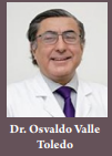
Review Article | Volume 1 | Issue 1 | JRS December 2021 | Page 39-43 | Osvaldo Valle Toledo. DOI: 10.13107/jrs.2021.v01.i01.021
Author: Osvaldo Valle Toledo [1]
[1] Department of Orthopedics and Traumatology, Ankle Foot Subspecialist, Ankle-Foot Team, MEDS Clinic, Santiago de Chile.
Address of Correspondence
Dr. Osvaldo Valle Toledo, MD,
Department of Orthopedics and Traumatology, Ankle Foot Subspecialist, Ankle-Foot Team, MEDS Clinic, Santiago de Chile.
E-mail: osvaldovalletoledo@yahoo.es
Abstract
Plantar fasciopathy is the most common cause of heel pain. It is a primarily degenerative and mechanical overuse pathology. The plantar fascia fulfills important biomechanical functions in the foot, being its “windlass” mechanism, the most important function in this regard, allowing the foot to act as a single and efficient motor unit during gait. Its clinical and imaging diagnosis is fully defined, being Baxter’s nerve entrapment neuropathy, its most significant differential diagnosis. The elongation exercises constitute the basic treatment, being the extracorporeal shock wave therapy of significant utility, amplified in its effects by the association with the referred therapeutic exercises.
Keywords: Plantar fasciitis, shock waves, fasciopathy.
References:
1. Rodríguez. Qué es la Fascia Plantar? 2015. Available from: https://lafisioterapia.net/que-es-la-fascia-plantar [Last accessed on 2021 Dec 12].
2. Buchanan BK, Kushner D. Plantar Fasciitis. Treasure Island, FL: StatPearls Publishing; 2021.
3. Monteagudo M, de Albornoz PM, Gutierrez B, Tabuenca J, Álvarez I. Plantar fasciopathy: A current concepts review. EFORT Open Rev 2018;3:485-93.
4. Pasapula C, Kiliyanpilakkil B, Khan DZ, Di Marco Barros R, Kim S, Ali AM, et al. Plantar fasciitis: Talonavicular instability/spring ligament failure as the driving force behind its histological pathogenesis. Foot (Edinb) 2021;46:101703.
5. Harutaichun P, Boonyong S, Pensri P. Differences in lower-extremity kinematics between the male military personnel with and without plantar fasciitis. Phys Ther Sport 2021;50:130-7.
6. Kirkpatrick J, Yassaie O, Mirjalili SA. The plantar calcaneal spur: A review of anatomy, histology, etiology and key associations. J Anat 2017;230:743-51.
7. Li J, Muehleman C. Anatomic relationship of heel spur to surrounding soft tissues: Greater variability than previously reported. Clin Anat 2007;20:950-5.
8. Díaz-Llopis IV. Despejando dudas sobre la fascitis plantar. XXIX Congreso de la Sociedad Valenciana de Medicina Física y Rehabilitación. Slides Presentation. Available from: https://svmefr.com/wp-content/uploads/2020/03/ISMAEL-DIAZ.pdf [Last accessed on 2021 Dec 12].
9. Forman WM, Green MA. The role of intrinsic musculature in the formation of inferior calcaneal exostoses. Clin Podiatr Med Surg 1990;7:217-23.
10. Acosta TB, Pérez YM, Tápanes SH, Cordero JE, Lottie AG, Aliaga B, et al. Bibliographic review. Rev Iberoamericana Fisiot Kinesiol 2008;11:26-31.
11. Finkenstaedt T, Siriwanarangsun P, Statum S, Biswas R, Anderson KE, Bae WC, et al. The calcaneal crescent in patients with and without plantar fasciitis: An ankle MRI study. AJR Am J Roentgenol 2018;211:1075-82.
12. Arnold MJ, Moody AL. Common running injuries: Evaluation and management. Am Fam Physician 2018;97:510-6.
13. Cotchett M, Lennecke A, Medica VG, Whittaker GA, Bonanno DR. The association between pain catastrophising and kinesiophobia with pain and function in people with plantar heel pain. Foot (Edinb) 2017;32:8-14.
14. Tschopp M, Brunner F. Diseases and overuse injuries of the lower extremities in long distance runners. Z Rheumatol 2017;76:443-50.
15. Baur D, Schwabl C, Kremser C, Taljanovic MS, Widmann G, Sconfienza LM, et al. Shear wave elastography of the plantar fascia: Comparison between patients with plantar fasciitis and healthy control subjects. J Clin Med 2021;10:2351.
16. Schillizzi G, Alviti F, D’Ercole C, Elia D, Agostini F, Mangone M, et al. Evaluation of plantar fasciopathy shear wave elastography: A comparison between patients and healthy subjects. J Ultrasound 2021;24:417-22..
17. Yucel I, Ozturan KE, Demiraran Y, Degirmenci E, Kaynak G. Comparison of high-dose extracorporeal shockwave therapy and intralesional corticosteroid injection in the treatment of plantar fasciitis. J Am Podiatr Med Assoc 2010;100:105-10.
18. Puttaswamaiah R, Chandran P. Degenerative plantar fasciitis: A review of current concepts. Foot 2007;17:3-9.
19. Buchbinder R, Ptasznik R, Gordon J, Buchanan J, Prabaharan V, Forbes A. Ultrasound-guided extracorporeal shock wave therapy for plantar fasciitis: A randomized controlled trial. JAMA 2002;288:1364-72.
20. Aqil A, Siddiqui MR, Solan M, Redfern DJ, Gulati V, Cobb JP. Extracorporeal shock wave therapy is effective in treating chronic plantar fasciitis: A meta-analysis of RCTs. Clin Orthop Relat Res 2013;471:3645-52..
21. Moya D, Ramón S, Schaden W, Wang CJ, Guiloff L, Cheng JH. The role of extracorporeal shockwave treatment in musculoskeletal disorders. J Bone Joint Surg Am 2018;100:251-63.
22. Sun J, Gao F, Wang Y, Sun W, Jiang B, Li Z. Extracorporeal shock wave therapy is effective in treating chronic plantar fasciitis: A meta-analysis of RCTs. Medicine (Baltimore) 2017;96:e6621.
23. Chang KV, Chen SY, Chen WS, Tu YK, Chien KL. Comparative effectiveness of focused shock wave therapy of different intensity levels and radial shock wave therapy for treating plantar fasciitis: A systematic review and network meta-analysis. Arch Phys Med Rehabil 2012;93:1259-68.
24. Greve JM, Grecco MV, Santos-Silva PR. Comparison of radial shockwaves and conventional physiotherapy for treating plantar fasciitis. Clinics (Sao Paulo) 2009;64:97-103.
25. Rompe JD, Meurer A, Nafe B, Hofmann A, Gerdesmeyer L. Repetitive low-energy shock wave application without local anesthesia is more efficient than repetitive low-energy shock wave application with local anesthesia in the treatment of chronic plantar fasciitis. J Orthop Res 2005;23:931-41.
26. Haddad S, Yavari P, Mozafari S, Farzinnia S, Mohammadsharifi G. Platelet-rich plasma or extracorporeal shockwave therapy for plantar fasciitis. Int J Burns Trauma 2021;11:1-8.
27. Llurda-Almuzara L, Labata-Lezaun N, Meca-Rivera T, Navarro-Santana MJ, Cleland JA, Fernández-de-Las-Peñas C, et al. Is dry needling effective for the management of plantar heel pain or plantar fasciitis? An updated systematic review and meta-analysis. Pain Med 2021;22:1630-41.
28. DiGiovanni BF, Nawoczenski DA, Lintal ME, Moore EA, Murray JC, Wilding GE, et al. Tissue-specific plantar fascia-stretching exercise enhances outcomes in patients with chronic heel pain. A prospective, randomized study. J Bone Joint Surg Am 2003;85:1270-7.
29. Avilés SG. Efectividad de las Ondas de Choque en la Fascitis Plantar. Revisión Sistemática. España: Alcalá la Real; 2017.
30. Schuitema D, Greve C, Postema K, Dekker R, Hijmans JM. Effectiveness of mechanical treatment for plantar fasciitis: A systematic review. J Sport Rehabil 2019;29:657-74.
31. Weil LS Jr., Roukis TS, Weil LS, Borrelli AH. Extracorporeal shock wave therapy for the treatment of chronic plantar fasciitis: Indications, protocol, intermediate results, and a comparison of results to fasciotomy. J Foot Ankle Surg 2002;41:166-72.
32. Maier M, Steinborn M, Schmitz C, Stäbler A, Köhler S, Pfahler M, et al. Extracorporeal shock wave application for chronic plantar fasciitis associated with heel spurs: Prediction of outcome by magnetic resonance imaging. J Rheumatol 2000;27:2455-62.
| How to Cite this article: Toledo OV | Plantar Fasciopathy, General Concepts, Shock Wave Treatment and Other Additional Therapeutic Consideration. | Journal of Regenerative Science | Dec 2021; 1(1): 39-43. |
[Full Text HTML] [Full Text PDF] [XML]
Fundamentals of the Treatment of Male Erectile Dysfunctions with Low Intensity Shockwaves
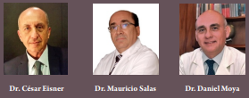
Review Article | Volume 1 | Issue 1 | JRS December 2021 | Page 26-29 | César Eisner, Mauricio Salas, Daniel Moya. DOI: 10.13107/jrs.2021.v01.i01.015
Author: César Eisner [1], Mauricio Salas [2], Daniel Moya [3]
[1] Shockwave Argentina, Buenos Aires, Argentina.
[2] Clínica Instituto de Urología y Sexología, Santiago de Chile, Chile.
[3] Department of Orthopaedics, Servicio de Ortopedia y Traumatología, Hospital Británico de Buenos Aires, Argentina.
Address of Correspondence
Dr. César Eisner, MD
Shockwave Argentina, Buenos Aires, Argentina.
E-mail: : info@shockwaveargentina.com
Abstract
Male erectile dysfunction (ED) is one of the most common problems among men worldwide. No single diagnostic method evaluates all the
variables of this complex condition. To achieve good therapeutic results, it is essential to base the treatment on an accurate diagnosis. Hemodynamic exploration by echo Doppler of the cavernous arteries, especially since the incorporation of intracavernous administration of vasoactive drugs, is a useful tool that allows the evaluation of erectile dysfunction in the arterial phase. It is also considered to be the choice in the assessment of the corporoveno-occlusive mechanism. Different treatment methods are used, being PDE5 (sildenafil and tadalafil), the treatment of the first choice in several conditions. The number of publications of low-intensity extracorporeal shockwave treatment (LI-ESWT) for ED has increased dramatically in recent years. Scientific evidence regarding the application of LI-ESWT for the treatment of erectile dysfunction is still controversial. Inclusion criteria of the studies and the wide variety of treatment protocols have been criticized. On the other hand, most of these studies report encouraging results with no short-term adverse effects, regardless of variation in LI-ESWT setup parameters or treatment protocols.
Keywords: Erectile dysfunction, Linear shock wave, Linear shock wave therapy, Shear wave elastography.
References:
1. Porst H. Review of the current status of low intensity extracorporeal shockwave therapy (Li-ESWT) in erectile dysfunction (ED), Peyronie’s disease (PD), and sexual rehabilitation after radical prostatectomy with special focus on technical aspects of the different marketed ESWT devices including personal experiences in 350 patients. Sex Med Rev 2021;9:93-122.
2. Clavijo RI, Kohn TP, Kohn JR, Ramasamy R. Effects of low-intensity extracorporeal shockwave therapy on erectile dysfunction: A systematic review and meta-analysis. J Sex Med 2017;14:27-35.
3. Dong L, Chang D, Zhang X, Li J, Yang F, Tan K, et al. Effect of low-intensity extracorporeal shock wave on the treatment of erectile dysfunction: A systematic review and meta-analysis. Am J Mens Health 2019;13:1557988319846749.
4. Lu Z, Lin G, Reed-Maldonado A, Wang C, Lee YC, Lue TF. Low-intensity extracorporeal shock wave treatment improves erectile function: A systematic review and meta-analysis. Eur Urol 2017;71:223-33.
5. Mo DS, Zhan XX, Shi HW, Cai HC, Meng J, Zhao J, et al. Efficacy and safety of low-intensity extracorporeal shock wave therapy in the treatment of ED: A meta-analysis of randomized controlled trials. Zhonghua Nan Ke Xue 2019;25:257-64.
6. Consensus Statement on ESWT Indications and Contraindications. Available from: https://www.shockwavetherapy.org/fileadmin/user_upload/dokumente/PDFs/Formulare/ISMST_consensus_statement_on_indications_and_contraindications_20161012_final.pdf [Last accessed on 2021 Oct 11].
7. Ondas de Choque en Medicina: La Nueva Frontera. Available from: https://onlat.net/?page_id=2491 [Last accessed on 2021 Oct 11].
8. Kessler A, Sollie S, Challacombe B, Briggs K, Van Hemelrijck M. The global prevalence of erectile dysfunction: A review. BJU Int 2019. Doi: 10.1111/bju.14813 Epub Ahead of Print.
9. Melman A, Rehman J. Pathophysiology of erectile dysfunction. Mol Urol 1999;3:87-102.
10. Jønler M, Moon T, Brannan W, Stone NN, Heisey D, Bruskewitz RC. The effect of age, ethnicity and geographical location on impotence and quality of life. Br J Urol 1995;75:651-5.
11. Huang SA, Lie JD. Phosphodiesterase-5 (PDE5) inhibitors in the management of erectile dysfunction. P T 2013;38:407-19.
12. Krzastek SC, Bopp J, Smith RP, Kovac JR. Recent advances in the understanding and management of erectile dysfunction. F1000Res. 2019;8:F1000 Faculty Rev-102.
13. Sokolakis I, Dimitriadis F, Psalla D, Karakiulakis G, Kalyvianakis D, Hatzichristou D. Effects of low-intensity shock wave therapy (LiST) on the erectile tissue of naturally aged rats. Int J Impot Res 2019;31:162-9.
14. Liu T, Shindel AW, Lin G, Lue TF. Cellular signaling pathways modulated by low-intensity extracorporeal shock wave therapy. Int J Impot Res 2019;31:170-6.
15. d’Agostino MC, Craig K, Tibalt E, Respizzi S. Shock wave as biological therapeutic tool: From mechanical stimulation to recovery and healing, through mechanotransduction. Int J Surg 2015;24 Pt B:147-53.
16. Moya D, Ramón S, Schaden W, Wang CJ, Guiloff L, Cheng JH. The role of extracorporeal shockwave treatment in musculoskeletal disorders. J Bone Joint Surg 2018;100:251-63.
17. Wang CJ. An overview of shock wave therapy in musculoskeletal disorders. Chang Gung Med J 2003;26:220-32.
18. Qiu X, Lin G, Xin Z, Ferretti L, Zhang H, Lue TF, et al. Effects of low-energy shockwave therapy on the erectile function and tissue of a diabetic rat model. J Sex Med 2013;10:738-46.
19. Rizk PJ, Krieger JR, Kohn TP, Pastuszak AW. Low-intensity shockwave therapy for erectile dysfunction. Sex Med Rev 2018;6:624-30.
20. Katz JE, Clavijo RI, Rizk P, Ramasamy R. The basic physics of waves, soundwaves, and shockwaves for erectile dysfunction. Sex Med Rev 2020;8:100-5.
21. Yee CH, Chan ES, Hou SS, Ng CF. Extracorporeal shockwave therapy in the treatment of erectile dysfunction: A prospective, randomized, double-blinded, placebo controlled study. Int J Urol 2014;21:1041-5.
22. Kalyvianakis D, Hatzichristou D. Low-intensity shockwave therapy improves hemodynamic parameters in patients with vasculogenic erectile dysfunction: A triplex ultrasonography-based sham-controlled trial. J Sex Med 2017;14:891-7.
23. Fojecki GL, Tiessen S, Osther PJ. Effect of low-energy linear shockwave therapy on erectile dysfunction-a double-blinded, sham-controlled, randomized clinical trial. J Sex Med 2017;14:106-12.
24. Olsen AB, Persiani M, Boie S, Hanna M, Lund L. Can low-intensity extracorporeal shockwave therapy improve erectile dysfunction? A prospective, randomized, double-blind, placebo-controlled study. Scand J Urol 2015;49:329-33.
25. Vardi Y, Appel B, Kilchevsky A, Gruenwald I. Does low intensity extracorporeal shock wave therapy have a physiological effect on erectile function? Short-term results of a randomized, double-blind, sham controlled study. J Urol 2012;187:1769-75.
26. Pelayo-Nieto M, Linden-Castro E, Alias-Melgar A, Espinosa-Pérez Grovas D, Carreño-de la Rosa F, Bertrand-Noriega F, et al. Linear shock wave therapy in the treatment of erectile dysfunction. Actas Urol Esp 2015;39:456-9.
27. Huang YP, Liu W, Liu YD, Zhang M, Xu SR, Lu MJ. Effect of low-intensity extracorporeal shockwave therapy on nocturnal penile tumescence and rigidity and penile haemodynamics. Andrologia 2020;52:e13745.
28. Domes T, Najafabadi BT, Roberts M, Campbell J, Flannigan R, Bach P, et al. Canadian urological association guideline: Erectile dysfunction. Can Urol Assoc J 2021;15:310-22.
29. Burnett AL, Nehra A, Breau RH, Culkin DJ, Faraday MM, Hakim LS, et al. Erectile dysfunction: AUA guideline. J Urol 2018;200:633-41.
30. Hackett G, Kirby M, Wylie K, Heald A, Ossei-Gerning N, Edwards D, et al. British society for sexual medicine guidelines on the management of erectile dysfunction in men-2017. J Sex Med 2018;15:430-57.
31. Hatzimouratidis K, Giuliano F, Moncada I, Muneer A, Salonia A, Verze P. EAU Guidelines on Erectile Dysfunction, Premature Ejaculation, Penile Curvature and Priapism. European Association of Urology; 2019. p. 23. Available from: https://uroweb.org/wp-content/uploads/EAU-guidelines-on-male-sexual-dysfunction-2019.pdf [Last accessed on 2021 Oct 11].
| How to Cite this article: Eisner C, Salas M, Moya D | Fundamentals of the Treatment of Male Erectile Dysfunctions with Low Intensity Shockwaves. | Journal of Regenerative Science | Dec 2021; 1(1): 26-29. |
[Full Text HTML] [Full Text PDF] [XML]
Non Invasive Phisical Physical Regenerative Therapies: Laser therapy, Mechanism of Action and Results
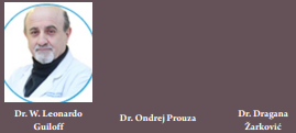
Review Article | Volume 1 | Issue 1 | JRS December 2021 | Page 21-25 | W. Leonardo Guiloff , Ondrej Prouza , Dragana Žarković. DOI: 10.13107/jrs.2021.v01.i01.013
Author: W. Leonardo Guiloff [1], Ondrej Prouza [2], Dragana Žarković [2]
[1] Department of Orthopedic Surgery of Davila Clinic, Santiago Chile, Past President Onlat-Achitoc, Santiago, Chile.
[2] Department of Anatomy and Biomechanics, Faculty of Physical Education and Sports, Charles
University, Prague, Czech Republic.
Address of Correspondence
Dr. W. Leonardo Guiloff, MD
Department of Orthopedic Surgery of Davila Clinic, Santiago Chile, Past President Onlat-Achitoc, Santiago, Chile.
E-mail: lguiloff@davila.cl
Abstract
Low-level laser therapy (LLLT) and high-intensity laser therapy (HILT) have emerged as a therapeutic alternative suitable for a wide range of medical conditions. The main advantage of high-intensity laser over LLLT is its ability to deliver a much higher dose in a shorter time while achieving deeper penetration into the affected tissue and producing a thermal effect. Although HILT, provides very satisfactory clinical results, more clinical research is require to justify its massive use.
Keywords: Low-level laser therapy, High-level laser therapy, Biostimulation, Phototherapy.
References:
1. Huang C, Holfeld J, Schaden W, Orgill D, Ogawa R. Mechanotherapy: Revisiting physical therapy and recruiting mechanobiology for a new era in medicine. Trends Mol Med 2013;19:555-64.
2. Moya D, Ramón S, Schaden W, Wang CJ, Guiloff L, Cheng JH. The role of extracorporeal shockwave treatment in musculoskeletal disorders. J Bone Joint Surg Am 2018;100:251-63.
3. Brañes J, Contreras HR, Cabello P, Antonic V, Guiloff LJ, Brañes M. Shoulder rotator cuff responses to extracorporeal shockwave therapy: Morphological and immunohistochemical analysis. Shoulder Elbow 2012;4:163-8.
4. Taylor N. Laser: The Inventor, the Nobel Laureate, and the Thirty Year Patent war. New York: Nick Taylor Simon Schuster; 2000.
5. Myers R, Dixon R. Who invented the laser: An analysis of the early patents. In: Historical Studies in the Physical and Biological Sciences. Vol. 34. California: University of California Press; 2003. p. 115-49.
6. Mester E. Risultati clinici di stimolazione laser e studi sperimentali circa il meccanismo di azione [Clinical results of laser stimulation and experimental studies on its mechanism of action]. Minerv Med. 1981;72:2195-9.
7. Huang YY, Chen AC, Carroll JD, Hamblin MR. Biphasic dose response in low level light therapy. Dose Response 2009;7:358-83.
8. Wickenheisser VA, Zywot EM, Rabjohns EM, Lee HH, Lawrence DS, Tarrant TK. Laser light therapy in inflammatory, musculoskeletal, and autoimmune disease. Curr Allergy Asthma Rep 2019;19:37.
9. Cotler HB, Chow RT, Hamblin MR, Carroll J. The use of low level laser therapy (LLLT) for musculoskeletal pain. MOJ Orthop Rheumatol 2015;2:00068.
10. Clijsen R, Brunner A, Barbero M, Clarys P, Taeymans J. Effects of low-level laser therapy on pain in patients with musculoskeletal disorders: A systematic review and meta-analysis. Eur J Phys Rehabil Med 2017;53:603-10.
11. Konstantinovic LM, Kanjuh ZM, Milovanovic AN, Cutovic MR, Djurovic AG, Savic VG, et al. Acute low back pain with radiculopathy: A double-blind, randomized, placebo-controlled study. Photomed Laser Surg 2010;28:553-60.
12. Konstantinovic LM, Cutovic MR, Milovanovic AN, Jovic SJ, Dragin AS, Letic MD, et al. Low-level laser therapy for acute neck pain with radiculopathy: A double-blind placebo-controlled randomized study. Pain Med 2010;11:1169-78.
13. Chow RT, Johnson MI, Lopes-Martins RA, Bjordal JM. Efficacy of low-level laser therapy in the management of neck pain: A systematic review and meta-analysis of randomised placebo or active-treatment controlled trials. Lancet 2009;374:1897-908.
14. Djavid GE, Mehrdad R, Ghasemi M, Hasan-Zadeh H, Sotoodeh-Manesh A, Pouryaghoub G. In chronic low back pain, low level laser therapy combined with exercise is more beneficial than exercise alone in the long term: A randomised trial. Aust J Physiother 2007;53:155-60.
15. Momenzadeh S, Kiabi FH, Moradkhani M, Moghadam MH. Low level laser therapy (LLLT) combined with physical exercise, a more effective treatment in low back pain. J Laser Med Sci 2012;3:67-70.
16. Tumilty S, Munn J, McDonough S, Hurley DA, Basford JR, Baxter GD. Low level laser treatment of tendinopathy: A systematic review with meta-analysis. Photomed Laser Surg 2010;28:3-16.
17. Bjordal JM, Lopes-Martins RA, Iversen VV. A randomised, placebo controlled trial of low level laser therapy for activated Achilles tendinitis with microdialysis measurement of peritendinous prostaglandin E2 concentrations. Br J Sports Med 2006;40:76-80; discussion 76-80.
18. Bjordal JM, Lopes-Martins RA, Joensen J, Couppe C, Ljunggren AE, Stergioulas A, et al. A systematic review with procedural assessments and meta-analysis of low level laser therapy in lateral elbow tendinopathy (tennis elbow). BMC Musculoskelet Disord 2008;9:75.
19. Bjordal JM, Couppé C, Chow RT, Tunér J, Ljunggren EA. A systematic review of low level laser therapy with location-specific doses for pain from chronic joint disorders. Aust J Physiother. 2003;49:107-16.
20. Tascioglu F, Armagan O, Tabak Y, Corapci I, Oner C. Low power laser treatment in patients with knee osteoarthritis. Swiss Med Wkly 2004;134:254-8.
21. Brosseau L, Welch V, Wells G, DeBie R, Gam A, Harman K, et al. Low level laser therapy (Classes I, II and III) for treating osteoarthritis. Cochrane Database Syst Rev 2004;3:CD002046.
22. Stasinopoulos DI, Johnson MI. Effectiveness of low-level laser therapy for lateral elbow tendinopathy. Photomed Laser Surg 2005;23:425-30.
23. Enwemeka CS, Parker JC, Dowdy DS, Harkness EE, Sanford LE, Woodruff LD. The efficacy of low-power lasers in tissue repair and pain control: A meta-analysis study. Photomed Laser Surg 2004;22:323-9.
24. Hopkins JT, McLoda TA, Seegmiller JG, Baxter GD. Low-level laser therapy facilitates superficial wound healing in humans: A triple-blind, sham-controlled study. J Athl Train 2004;39:223-9.
25. Fukuda VO, Fukuda TY, Guimarães M, Shiwa S, de Lima Bdel C, Martins RÁ, et al. Short-term Eefficacy of low-level laser therapy in patients with knee osteoarthritis: A randomized placebo-controlled, double-blind clinical trial. Rev Bras Ortop 2015;46:526-33.
26. Conti PC. Low level laser therapy in the treatment of temporomandibular disorders (TMD): A double-blind pilot study. Cranio 1997;15:144-9.
27. Brosseau L, Robinson V, Wells G, Debie R, Gam A, Harman K, et al. Low level laser therapy (Classes I, II and III) for treating rheumatoid arthritis. Cochrane Database Syst Rev 2005;4:CD002049.
28. Stiglić-Rogoznica N, Stamenković D, Frlan-Vrgoc L, Avancini-Dobrović V, Vrbanić TS. Analgesic effect of high intensity laser therapy in knee osteoarthritis. Coll Antropol 2011;35 Suppl 2:183-5.
29. Prochazka M. “Class IV Laser in Noninvasive Laser Therapy.” Laser Partner; 2006. Available from: https://www.laserpartner.org/lasp/web/en/2003/0069html [Last access date 2006].
30. Bettencourt F. Effects of class IV laser in knee osteoarthritis: A randomized control trial. J Arthritis 2020;9:1-289.
31. Vescovi P, Merigo E, Manfredi M, Meleti M, Fornaini C, Bonanini M, et al. Nd: YAG laser biostimulation in the treatment of bisphosphonate-associated osteonecrosis of the jaw: Clinical experience in 28 cases. Photomed Laser Surg 2008;26:37-46.
32. Santamato A, Solfrizzi V, Panza F, Tondi G, Frisardi V, Leggin BG, et al. Short-term effects of high-intensity laser therapy versus ultrasound therapy in the treatment of people with subacromial impingement syndrome: A randomized clinical trial. Phys Ther 2009;89:643-52.
33. Wyszyńska J, Bal-Bocheńska M. Efficacy of high-intensity laser therapy in treating knee osteoarthritis: A first systematic review. Photomed Laser Surg 2018;36:343-53.
34. Graudenz K, Raulin C. Von einsteins quantentheorie zur modernen lasertherapie: Historie des lasers in der dermatologie und ästhetischen medizin. Hautarzt 2003;7:575-82.
35. Kojevnikov A. Nikolay Gennadiyevich Basov. Encyclopedia Britannica; 2021. Available from: https://www.britannica.com/biography/Nikolay-Basov [Last accessed on 2021 Aug 2].
36. Charles H. Townes Biographical. NobelPrize.org. Nobel Prize Outreach AB 2021. Sun. 31 Oct; 2021. Available from: https://www.nobelprize.org/prizes/physics/1964/townes/biographical [Last accessed on 2021 Aug 2].
37. Schawlow AL. Schawlow Biographical. NobelPrize.org. Nobel Prize Outreach AB 2021. Sun. 31 Oct; 2021. Available from: https://www.nobelprize.org/prizes/physics/1981/schawlow/biographical [Last accessed on 2021 Aug 2].
38. Maiman TH. Stimulated optical radiation in ruby. Nature 1960;187:493-4.
39. Sommer AP, Pinheiro AL, Mester AR, Franke RP, Whelan HT. Biostimulatory windows in low-intensity laser activation: Lasers, scanners, and NASA’s light-emitting diode array system. J Clin Laser Med Surg 2001;19:29-33.
40. Nouri K, Ballard CJ. Laser therapy for acne. Clin Dermatol 2006;24:26-32.
41. Evans DH, Abrahamse H. Efficacy of three different laser wavelengths for in vitro wound healing. Photodermatol Photoimmunol Photomed 2008;24:199-210.
42. Whelan HT, Smits RL Jr., Buchman EV, Whelan NT, Turner SG, Margolis DA, et al. Effect of NASA light-emitting diode irradiation on wound healing. J Clin Laser Med Surg 2001;19:305-14.
43. Peplow PV, Chung TY, Baxter GD. Laser photobiomodulation of wound healing: A review of experimental studies in mouse and rat animal models. Photomed Laser Surg 2010;28:291-325.
44. Conlan MJ, Rapley JW, Cobb CM. Biostimulation of wound healing by low-energy laser irradiation. A review. J Clin Periodontol 1996;23:492-6.
45. Woodruff LD, Bounkeo JM, Brannon WM, Dawes KS, Barham CD, Waddell DL, et al. The efficacy of laser therapy in wound repair: A meta-analysis of the literature. Photomed Laser Surg 2004;22:241-7.
46. Dundar U, Turkmen U, Toktas H, Solak O, Ulasli AM. Effect of high-intensity laser therapy in the management of myofascial pain syndrome of the trapezius: A double-blind, placebo-controlled study. Lasers Med Sci 2015;30:325-32.
47. Kneebone WJ, Crna DC, DIHom CN. Practical applications of low level laser therapy. Pract Pain Manag 2006;6:34-40.
48. Marshall RP, Vlková K. Spectral dependence of laser light on light-tissue interactions and its influence on laser therapy: An experimental study. Insights Biomed 2020;5:1.
49. Baack HO. Comparison of energy spread homogeneity in automated and manual class 4 laser therapy. Res Rev 2020;9:1-9.
50. Khalkhal E, Razzaghi M, Rostami-Nejad M, Rezaei-Tavirani M, Heidari Beigvand H, Tavirani MR. Evaluation of laser effects on the human body after laser therapy. J Lasers Med Sci 2020;11:91-7.
51. Tseng SH, Bargo P, Durkin A, Kollias N. Chromophore concentrations, absorption and scattering properties of human skin in-vivo. Opt Express 2009;17:14599-617.
52. Chen CH, Tsai JL, Wang YH, Lee CL, Chen JK, Huang MH. Low-level laser irradiation promotes cell proliferation and mRNA expression of Type I collagen and decorin in porcine Achilles tendon fibroblasts in vitro. J Orthop Res 2009;27:646-50.
53. Karu TI, Hode L. Ten Lectures on Basic Science of Laser Phototherapy. Grängesberg: Prima Books; 2007.
54. Mester A, Mester A. The history of photobiomodulation: Endre mester (1903-1984). Photomed Laser Surg 2017;35:393-4.
55. Peplow PV, Chung TY, Baxter GD. Laser photobiomodulation of proliferation of cells in culture: A review of human and animal studies. Photomed Laser Surg 2010;28 Suppl 1:S3-40.
56. Karu T. Mitochondrial mechanisms of photobiomodulation in context of new data about multiple roles of ATP. Photomed Laser Surg 2010;28:159-60.
57. Holden PK, Li C, Da Costa V, Sun CH, Bryant SV, Gardiner DM, et al. The effects of laser irradiation of cartilage on chondrocyte gene expression and the collagen matrix. Lasers Surg Med 2009;41:487-91.
58. Chen CH, Hung HS, Hsu SH. Low-energy laser irradiation increases endothelial cell proliferation, migration, and eNOS gene expression possibly via PI3K signal pathway. Lasers Surg Med 2008;40:46-54.
59. Zati A, Desando G, Cavallo C, Buda R, Giannini S, Fortuna D, et al. Treatment of human cartilage defects by means of Nd: YAG laser therapy. J Biol Regul Homeost Agents 2012;26:701-11.
60. Peat FJ, Colbath AC, Bentsen LM, Goodrich LR, King MR. In vitro effects of high-intensity laser photobiomodulation on equine bone marrow-derived mesenchymal stem cell viability and cytokine expression. Photomed Laser Surg 2018;36:83-91.
61. Bjordal JM, Lopes-Martins RA, Joensen J, Iversen VV. The anti-inflammatory mechanism of low level laser therapy and its relevance for clinical use in physiotherapy. Phys Ther Rev 2010;15:286-293.
62. Walker J. Relief from chronic pain by low power laser irradiation. Neurosci Lett 1983;43:339-44.
63. Fiore P, Panza F, Cassatella G, Russo A, Frisardi V, Solfrizzi V, et al. Short-term effects of high-intensity laser therapy versus ultrasound therapy in the treatment of low back pain: A randomized controlled trial. Eur J Phys Rehabil Med 2011;47:367-73.
64. Bálint G, Barabás K, Zeitler Z, Bakos J, Kékesi KA, Pethes A, et al. Ex vivo soft-laser treatment inhibits the synovial expression of vimentin and α-enolase, potential autoantigens in rheumatoid arthritis. Phys Ther 2011;91:665-74.
65. Paradiz SG, Guiloff L, Aroca MB. HILT (High Intensity Laser Therapy) HILT and RSWT (Radial Schockwave Treatment): Pain and Pacient Satisfaction Evaluation of 100 Patients. Available from: https://www.shockwavetherapy.org/fileadmin/user_upload/dokumente/PDFs/Abstracts/abstracts-ismst-congress-18-mendoza-2015.pdf [Last accessed on 2021 Aug 2].
66. Coombes BK, Bisset L, Vicenzino B. A new integrative model of lateral epicondylalgia. Br J Sports Med 2009;43:252-8.
67. Armagan O, Tascioglu F, Ekim A, Oner C. Long-term efficacy of low level laser therapy in women with fibromyalgia: A placebo-controlled study. J Back Musculoskelet Rehabil 2006;19:135-40.
68. Jovicić M, Konstantinović L, Lazović M, Jovicić V. Clinical and functional evaluation of patients with acute low back pain and radiculopathy treated with different energy doses of low level laser therapy. Vojnosanit Pregl 2012;69:656-62.
69. Rochkind S, Geuna S, Shainberg A. Chapter 25: Phototherapy in peripheral nerve injury: Effects on muscle preservation and nerve regeneration. Int Rev Neurobiol 2009;87:445-64.
70. Kawecki M, Bernad-Wiśniewska T, Sakiel S, Nowak M, Andriessen A. Laser in the treatment of hypertrophic burn scars. Int Wound J 2008;5:87-97.
71. Marotti J, Sperandio FF, Fregnani ER, Aranha AC, de Freitas PM, de Eduardo CP. High-intensity laser and photodynamic therapy as a treatment for recurrent herpes labialis. Photomed Laser Surg 2010;28:439-44.
72. Kozarev J, Vizintin Z. Novel laser therapy in treatment of onychomycosis. J Laser Health Acad 2010;1:1-8..
73. Hochman LG. Laser treatment of onychomycosis using a novel 0.65-millisecond pulsed Nd: YAG 1064-nm laser. J Cosmet Laser Ther 2011;13:2-5.
| How to Cite this article: Guiloff WL, Prouza O, Žarković D | Non-Invasive Physical Regenerative Therapies: Laser therapy, Mechanism of Action and Results. | Journal of Regenerative Science | Dec 2021; 1(1): 21-25. |
[Full Text HTML] [Full Text PDF] [XML]
The Sports, Ultrasound, Biologics, and Arthroscopy Protocol in the New Era of Orthopaedic Sports Injuries Treatments
Review Article | Volume 1 | Issue 1 | JRS December 2021 | Page 16-20 | Bernáldez Domínguez Pedro, Dallo Lazzarini Ignacio. DOI: 10.13107/jrs.2021.v01.i01.011
Author: Bernáldez Domínguez Pedro [1], Dallo Lazzarini Ignacio [1]
[1] Department of Orthopaedic Surgery and Sports Medicine, SportMe Medical Center, Unit of Biological Therapies and Ultrasounds, Seville, Spain.
Address of Correspondence
Dr. Bernáldez Domínguez Pedro, MD. PhD
Tabladilla, 2, 41013, Seville, Spain.
E-mail: pedrobernaldez@gmail.com
Abstract
In the new era of sports traumatology, the union of anatomical, biomechanical, and functional knowledge, together with an adequate clinical examination and complemented with ultrasound studies, arthroscopic surgery, and conventional surgery, makes us understand the pathology, in a new and modern way, of the locomotor system, such as the muscle, tendon, ligament, menisci, capsule, synovial membrane, as well as bone and cartilage pathologies. Biological therapies have shown a good result for soft tissue in chronic pathology that can be applied in an ultrasound guided manner to treat tendinopathy of the Achilles, patellar, and quadriceps tendons, also at the elbow and shoulder level. It is striking to highlight the good results of this biological therapy with platelet-rich plasma for degenerative joint diseases in patients with moderate osteoarthritis. In cases in which conservative or biological therapies have not had their effect, we will generally indicate surgery, in most cases arthroscopically if it is joint pathology. This indication will be mandatory, especially in joint instability cases where we will require stabilizing surgery. We emphasize the importance of multidisciplinary teams where there must be a sports doctor, a sports traumatologist, a physiotherapist, a functional trainer, a podiatrist, biomechanics specialist, and other professionals that surround the athlete, such as the nutritionist, the psychologist so that the athlete has comprehensive assistance and is always well cared for. Together, these concepts make a personalized approach named the Sports, Ultrasound, Biologics, and Arthroscopy protocol to improve clinical results, shorten recovery times, and considerably reduce healthcare costs.
Keywords: Sports, Ultrasound, Biologics, Arthroscopy protocol, Sports medicine, Ultrasound-guided therapies, Biological therapies, Arthroscopy.
References:
1. Lind M, Seil R, Dejour D, Becker R, Menetrey J, Ross M. Creation of a specialist core curriculum for the European Society for Sports traumatology, Knee surgery and Arthroscopy (ESSKA). Knee Surg Sports Traumatol Arthrosc 2020;28:3066-79.
2. Centeno C. In: Aldridge K, editor. Orthopedics 2.0: How Regenerative Medicine and Interventional Orthopedics will Change Everything. RHIA; 2018.
3. Daniels EW, Cole D, Jacobs B, Phillips SF. Existing evidence on ultrasound-guided injections in sports medicine. Orthop J Sports Med 2018;6:2325967118756576.
4. Hoeber S, Aly AR, Ashworth N, Rajasekaran S. Ultrasound-guided hip joint injections are more accurate than landmark-guided injections: A systematic review and meta-analysis. Br J Sports Med 2016;50:392-6.
5. Peck E, Jelsing E, Onishi K. Advanced ultrasound-guided interventions for tendinopathy. Phys Med Rehabil Clin N Am 2016;27:733-48.
6. Domínguez B. Martos AT. El ecógrafo: El fonendo del Traumatólogo: Utilidad diagnostica y terapéutica. Rev S Traum Ort 2017;34:17-26.
7. Dallo I, Chahla J, Mitchell JJ, Pascual-Garrido C, Feagin JA, LaPrade RF. Biologic approaches for the treatment of partial tears of the anterior cruciate ligament: A current concepts review. Orthop J Sports Med 2017;5:2325967116681724.
8. Kon E, Di Matteo B, Delgado D, Cole BJ, Dorotei A, Dragoo JL, et al. Platelet-rich plasma for the treatment of knee osteoarthritis: An expert opinion and proposal for a novel classification and coding system. Expert Opin Biol Ther 2020;20:1447-60.
9. Le AD, Enweze L, DeBaun MR, Dragoo JL. Current clinical recommendations for use of platelet-rich plasma. Curr Rev Musculoskelet Med 2018;11:624-34.
10. Piuzzi NS, Khlopas A, Newman JM, Ng M, Roche M, Husni ME, et al. Bone marrow cellular therapies: Novel therapy for knee osteoarthritis. J Knee Surg 2018;31:22-6.
11. Gobbi A, Whyte GP. Long-term clinical outcomes of one-stage cartilage repair in the knee with hyaluronic acid-based scaffold embedded with mesenchymal stem cells sourced from bone marrow aspirate concentrate. Am J Sports Med 2019;47:1621-8.
12. Gobbi A, Karnatzikos G, Scotti C, Mahajan V, Mazzucco L, Grigolo B. One-Step cartilage repair with bone marrow aspirate concentrated cells and collagen matrix in full-thickness knee cartilage lesions: Results at 2-year follow-up. Cartilage 2011;2:286-99.
13. Dallo I, Morales M, Gobbi A. Platelets and Adipose Stroma Combined for the Treatment of the Arthritic Knee. Arthroscopy Techniques; November 2021.
14. Gobbi A, Dallo I, Rogers C, Striano RD, Mautner K, Bowers R, et al. Two-year clinical outcomes of autologous microfragmented adipose tissue in elderly patients with knee osteoarthritis: A multi-centric, international study. Int Orthop 2021;45:1179-88.
15. Jackson RW. A history of arthroscopy. Arthroscopy 2010;26:91-103.
16. Perets I, Rybalko D, Mu BH, Friedman A, Morgenstern DR, Domb BG. Hip Arthroscopy: Extra-articular Procedures. Hip Int 2019;29:346-54.
17. Chloros GD, Shen J, Mahirogullari M, Wiesler ER. Wrist arthroscopy. J Surg Orthop Adv 2007;16:49-61.
18. DiFelice GS, van der List JP. Clinical outcomes of arthroscopic primary repair of proximal anterior cruciate ligament tears are maintained at mid-term follow-up. Arthroscopy 2018;34:1085-93.
19. van der List JP, DiFelice GS. Primary repair of the anterior cruciate ligament: A paradigm shift. Surgeon 2017;15:161-8.

| How to Cite this article: Pedro BD, Ignacio DL | The Sports, Ultrasound, Biologics, and Arthroscopy Protocol in the New Era of Orthopaedic Sports Injuries Treatments. | Journal of Regenerative Science | Dec 2021; 1(1): 16-20. |
