History of Shock Waves and Radial Pressure Waves From Newton to Our Times
Review Article | Volume 3 | Issue 1 | JRS Jun – June 2023 | Page 09-14 | Daniel Moya, Achim M. Loske, Paul Hobrough , Carla Moya.
DOI: 10.13107/jrs.2023.v03.i01.70
Author: Daniel Moya [1], Achim M. Loske [2], Paul Hobrough [3], Carla Moya [4]
[1] Department of Orthopaedics, Hospital Británico de Buenos Aires, Argentina,
[2] Centro de Física Aplicada y Tecnología Avanzada, Universidad Nacional Autónoma de México, Juriquilla, Querétaro, México,,
[3] The University of Northumbria – Newcastle upon Tyne, UK,
[4] Instituto Tecnológico de Buenos Aires, Argentina.
Address of Correspondence
Dr. Daniel Moya,
Department of Orthopaedics, Hospital Británico de Buenos Aires, Argentina.
E-mail: drdanielmoya@yahoo.com.ar
Abstract
Although shock waves have been present in nature since its origins, current knowledge took many centuries of study and research. The history of the development of the use of mechanical waves for therapeutic purposes has been a long process in which scientists from many countries contributed. The physical knowledge of waves, the development of generation sources, the first applications in kidney stones and the discovery of their biological impact have been milestones in this long process. The aim of this publication is to highlight previous discoveries that have not been described when analyzing the development of shock waves as a therapeutic tool.
Keywords: Shock waves, Radial pressure waves, History.
References:
- Krehl P. History of shock waves. In: Ben-Dor G, Igra O, Elperin T, editors. Handbook of Shock Waves. Vol. 1. United States: Academic Press; 2001. p. 1-142.
- Newton I. Philosophiae naturalis principia mathematica; 2009. Available from: https://www.gutenberg.org/files/28233/28233-pdf.pdf Last accessed 29 Jan 2023
- Goriely A, McMillen T. Shape of a cracking whip. Phys Rev Lett 2002;88:244301.
- Lennauchfilm-Studio. He tamed the lightning. Science and technology film; 1957. Available from: https://www.calameo.com/read/00103816437ddb2395bc0
- Bekaev AA, Sokovikov VK, Strokov PI. Electrohydraulic devices based on the yutkin effect. Russ Eng Res 2014;34:620-3.
- Malinaric R, Mantica G, Martini M, Balzarini F, Mariano F, Marchi G, et al. The lifetime history of the first Italian public extra-corporeal shock wave lithotripsy (ESWL) lithotripter as a mirror of the evolution of endourology over the last decade. Int J Environ Res Public Health 2023;20:4127.
- Loske AM. Medical and biomedical applications of shock waves. Cham, Switzerland: Springer International; 2017. p. 55.
- Coleman AJ, Saunders JE, Crum LA, Dyson M. Acoustic cavitation generated by an extracorporeal shockwave lithotripter. Ultrasound Med Biol 1987;13:69-76.
- Delius M, Jordan M, Eizenhoefer H, Marlinghaus E, Heine G, Liebich HG, et al. Biological effects of shock waves: Kidney haemorrhage by shock waves in dogs–administration rate dependence. Ultrasound Med Biol 1988;14:689-94.
- Schelling G, Delius M, Gschwender M, Grafe P, Gambihler S. Extracorporeal shock waves stimulate frog sciatic nerves indirectly via a cavitation-mediated mechanism. Biophys J 1994;66:133-40.
- Angstman NB, Kiessling MC, Frank HG, Schmitz C. High interindividual variability in dose-dependent reduction in speed of movement after exposing elegans to shock waves. Front Behav Neurosci 2015;9:12.
- Császár NB, Angstman NB, Milz S, Sprecher CM, Kobel P, Farhat M, et al. Radial shock wave devices generate cavitation. PLoS One 2015;10:e0140541.
- Hazen RH, Boctor N, Brandes JA, Cody GD, Hemley RJ, Sharma A, et al. High pressure and the origin of life. J Phy Condens Matter 2022;14:11489.
- Kaminski A, Fedorchak GR, Lammerding J. The cellular mastermind(?)-mechanotransduction and the nucleus. Prog Mol Biol Transl Sci 2014;126:157-203.
- Mayhew E. The Invisible Killer of World War I: Blast Injury. Available from: https://www.knowitwall.com/episodes/the-invisible-killer-of-world-war-i-blast-injury Last accessed 29 Jan 2023
- Shoja MM, Tubbs RS. Augusta Déjerine-Klumpke: The first female neuroanatomist. Clin Anat 2007;20:585-7.
- Thiel M. Application of shock waves in medicine. Clin Orthop 2001;387:18-21.
- Wang CJ. An overview of shock wave therapy in musculoskeletal disorders. Chang Gung Med J 2003;26:220-32.
- Scott TE, Kirkman E, Haque M, Gibb IE, Mahoney P, Hardman JG. Primary blast lung injury-a review. Br J Anaesth 2017;118:311-6.
- Mackenzie I, Tunnicliffe B, Clasper JC, Mahoney P, Kirkman E. What the intensive care doctor needs to know about blast-related lung injury. J Intensive Care Soc 2013;14:303-12.
- Horrocks CL. Blast injuries: Biophysics, pathophysiology and management principles. J R Army Med Corps 2001;147:28-40.
- Yang C, Dong-Hai Z, Ling-Ying L, Yong-Hui Y, Yang W, Li-Wei Z, et al. Author correction: Simulation of blast lung injury induced by shock waves of five distances based on finite element modeling of a three-dimensional rat. Sci Rep 2019;9:3440.
- Delius M, Enders G, Heine G, Stark J, Remberger K, Brendel W. Biological effects of shock waves: Lung hemorrhage by shock waves in dogs–pressure dependence. Ultrasound Med Biol 1987;13:61-7.
- Nouri-Majalan N, Masoumi R, Halvani A, Moghaddasi S. Lung contusion and cavitation with exudative plural effusion following extracorporeal shock wave lithotripsy in an adult: A case report. J Med Case Rep 2010;4:293.
- Yi JH, Wang D, Chen H, Li ZS, Hu LH. Lung contusion after extracorporeal shock wave lithotripsy for pancreatic stones: A case report. Medicine (Baltimore) 2022;101:e30063.
- Chaussy C, Eisenberger F, Forssmann B. Extracorporeal shockwave lithotripsy (ESWL): A chronology. J Endourol 2007;21:1249-53.
- Keller EX, Proietti S, Talso M, Emiliani E, Ploumidis A, Mantica G, et al. Prone versus supine percutaneous nephrolithotomy: A systematic review and meta-analysis of current literature. Minerva Urol. Nephrol 2021;73:50-8.
- Farr JB. Frank rieber: Obscure genius (Part 1). Lead Edge 2008;27:613-8.
- Tefekli A, Cezayirli F. The history of urinary stones: In parallel with civilization. ScientificWorldJournal 2013;2013:423964.
- Mulvaney WP. Attempted disintegration of calculi by ultrasonic vibrations. J Urol 1953;70:704-7.
- European Museum of Urology (EMU). Available from: https://history.uroweb.org/history-of-urology/early-rologicalinterventions/lithotripsy/electrohydraulic-lithotripsy Last accessed 29 Jan 2023
- Tufano A, Frisenda M, Rossi A, Viscuso P, Mantica G, Bove P, et al. External validation of Resorlu-Unsal stone score in predicting outcomes after retrograde intrarenal surgery. Experience from a single institution. Arch Ital Urol Androl 2022;94:311-4.
- Haeusler E, Kiefer W. Anregung von Stoßwellen in Flüssigkeiten durch Hochgeschwindigkeitswassertropfen. [Excitation of shock waves in liquids by high-velocity water droplets] Verhandl. DPG (VI) 1971;6: K 36. (VI) 1971;6:K 36.
- Eisenberger F, Chaussy CH, Wanner K. Extrakorporale anwen-dung von hochenergetischen stosswellen: Ein neuer aspekt in derbehandlung des harnsteinleidens. [Extracorporeal application of high-energy shock waves: A new aspect in the treatment of urinary stone disease.] Akt Urol 1977;8:3-15.
- Chaussy CH, Schmiedt E, Forssmann B, Brendel W. Contact free renal stone destruction by means of shock waves. Eur Surg Res 1979;11:36.
- Chaussy C, Eisenberger F, Wanner K. Die implantation humaner nierensteine: Ein einfachesexperimentelles steinmodell. [The implantation of human kidney stones: A simple experimental step model] Urol A 1977;16:35-8.
- Forssmann B, Hepp W, Chaussy C, Jocham D, Schmiedt E, Brendel W. Prototyp für die klinischeanwendung der berührungsfreien nierensteinzertrümmerung durch stosswellen. [Prototype for the clinical application of non-contact urinary stone destruction by shock waves.] Biomed Tech 1980;25:414-6.
- Lohrer H, Nauck T, Korakakis V, Malliaropoulos N. Historical ESWT paradigms are overcome: A narrative review. Biomed Res Int 2016;2016:3850461.
- Chaussy C, Brendel W, Schmiedt E. Extracorporeally induced destruction of kidney stones by shock waves. Lancet 1980;2:1265-8.
- Haupt G. Use of extracorporeal shock waves in the treatment of pseudarthrosis, tendinopathy and other orthopedic diseases. J Urol 1997;158:4-11.
- Pearson OM, Lieberman DE. The aging of Wolff‘s “law”: Ontogeny and responses to mechanical loading in cortical bone. Am J Phys Anthropol 2004;Suppl 39:63-99.
- Thompson DA. On Growth and Form. United Kingdom: Cambridge University Press; 1917.
- Thompson DA. In: Bonner JT, editor. On Growth and Form. United Kingdom: Cambridge University Press; 1992.
- Garduño FS, Sánchez JL. Las transformaciones de Thompson y las formas biológicas. [Thompson transformations and biological forms] Ciencias 2017;126, 42-53.
- Iskratsch T, Wolfenson H, Sheetz MP. Appreciating force and shape-the rise of mechanotransduction in cell biology. Nat Rev Mol Cell Biol 2014;15:825-33.
- Lim CT, Bershadsky A, Sheetz MP. Mechanobiology. J R Soc Interface 2010;7 Suppl 3:S291-3.
- Fung YC. Biomechanics: Mechanical properties of living tissues. New York: Springer; 1993. p. 165-219.
- Martino F, Perestrelo AR, Vinarský V, Pagliari S, Forte G. Cellular mechanotransduction: From tension to function. Front Physiol 2018;9:824.
- Jaalouk DE, Lammerding J. Mechanotransduction gone awry. Nat Rev Mol Cell Biol 2009;10:63-73.
- Iqbal J, Zaidi M. Molecular regulation of mechanotransduction. Biochem Biophys Res Commun 2005;328:751-5.
- Graff J, Pastor J, Richter KD. Effect of high energy shock waves on bony tissue. Urol Res 1988;16:252-8.
- Haupt G, Chvapil M. Effect of shock waves on the healing of partial-thickness wounds in piglets. J Surg Res 1990;49:45-8.
- Haupt G, Haupt A, Chvapil M, Drach GW, Senge TH. Wound and fracture healing: n ew indication for extracorporealshock waves? J Endourol Suppl 1990;4:S54, abstract A-4.
- Haupt G, Haupt A, Ekkernkamp A, Gerety B, Chvapil M. Influence of shock waves on fracture healing. Urology 1992;39:529-532. DOI: 10.1016/0090-4295(92)90009-l.
- Wang FS, Wang CJ, Huang HC, Chung H, Chen RF, Yang KD. Physical shock wave mediates membrane hyperpolarization and Ras activation for osteogenesis in human bone marrow stromal cells. Biochem Biophys Res Commun 2001;287:648-55.
- Wang CJ, Huang HY, Pai CH. Shock wave-enhanced neovascularization at the tendon-bone-junction. An experiment in dogs. J Foot Ankle Surg 2002;41:16-22.
- Brañes J, Contreras HR, Cabello P, Antonic V, Guiloff LJ, Brañes M. Shoulder rotator cuff responses to extracorporeal shockwave therapy: Morphological and immunohistochemical analysis. Shoulder Elbow 2012;4:163-8.
- Gollmann-Tepeköylü C, Pölzl L, Graber M, Hirsch J, Nägele F, Lobenwein D, et al. miR-19a-3p containing exosomes improve function of ischaemic myocardium upon shock wave therapy. Cardiovasc Res 2020;116:1226-36.
- Gloeck TM. Effects of radial extracorporeal shock waves on healthy bone tissue in rabbit models. Complete copy of the dissertation approved by the Faculty of Medicine of the Technical University of Munich for obtaining the academic degree of Doctor of Medicine. Available from: https://d-nb.info/994118236/34 Last accessed 29 Jan 2023
- Horn CP. Effect of extracorporeal high-energy shock waves on bacteria and their interaction with antibiotics. Complete copy of the dissertation approved by the Faculty of Medicine of the Technical University of Munich for obtaining the academic degree of Doctor of Medicine. Available from: https://mediatum.ub.tum.de/doc/620254/620254.pdf Last accessed 29 Jan 2023
- Karpman RR, Magee FP, Gruen TW, Mobley T. Work-in-progress #1. The lithotriptor and its potential use in the revision of total hip arthroplasty. Orthop Rev 1987;16:38-42.
- Karpman RR, Magee FP, Gruen TW, Mobley T. The Lithotriptor and its potential use in the revision of total hip arthroplasty. Clin Orthop 2001;387:4-7.
- Valchanov V, Michailov P, Patrashkov T. New Possibilities of HM-3 lithotriptor for treatment of disturbed bone union. Read at World Congress on ESWL and Endourology, Kyoto, Japan; 1989.
- Valchanov VD, Michailov P. High energy shock waves in the treatment of delayed and nonunion fractures. Int Orthop 1991;15:181-4.
- Valchanov VD. Method and Aparattus for Medical Treatment of Pathological State of Bones. US Patent 4979501; 1990. Available from: https://image-ppubs.uspto.gov/dirsearch-public/print/downloadPdf/4979501 Last accessed 29 Jan 2023
- Haist J. Die osteorestauration via stobwellenanwendung. Eine neue moglichkeit zur therapie der gestorten knochernen konsolidierung. [Osteorestoration via shock wave application. A new possibility for the therapy of disturbed bony consolidation. In: Chaussy C, Eisenberger F, Jocham D Wilbert D, editors. Die StoOwelle-Forschung und Klinik. Tubingen: Attempto Verlag; 1995. p. 157.
- Dahmen GP, Meiss L, Nam VC, Franke R, Gonchars V. Extrakorporale stonwellentherapie (ESWT) im knochennahen weichteilbereich an der schulter. [Extracorporeal sound wave therapy (ESWT) in the soft tissue area close to the bone on the shoulder.] Erste therapieergebnisse. Ext Orthopaed 1992;15:25.
- Dahmen GP, Nam VC, Meiss L. Extrakorporale Stoßwellentherapie (ESWT) zur Behandlung von Knochennahen Weichteilschmerzen. Indikation, Technik und Vorläufige Ergebnisse. Konsensus Workshop der Deutschen Gesellschaft für Stoßwellenlithotripsie. [Indication, Technique and Preliminary Results. Consensus workshop of the German Society for Shockwave Lithotripsy.] Tübingen: Attempto-Verlag; 1993. p. 143-8.
- Dahmen GP, Franke R, Gonchars V, Poppe S, Lentrodt S, Lichtenberger S, et al. Die behandlung knochennaher weichteilschmerzen mit extrakorporaler stosswellentherapie (ESWT), Indikation, Technik und bisherige therapie. [The treatment of soft tissue pain close to the bone with extracorporeal shock wave therapy (ESWT), indication, technique and previous therapy.] In: Chaussy C, Eisenberger F, Jochum D, Wilbert D, editors. Die Stoßwelle-Forschung und Klinik. Tübingen, Germany: Attempo Verlag; 1995. p. 175-86.
- Loew M, Jurgowski W. Initial experiences with extracorporeal shockwave lithotripsy (ESWL) in treatment of tendinosis calcarea of the shoulder. Z Orthop Ihre Grenzgeb 1993;131:470-3.
- Loew M, Jurgowski W, Mau H, Perlick L, Kuszniercak D. Die wirkung extrakorporal erzeugter hochenergetischer storwellen auf den klinischen, rontgenologischen undhistologischen verlauf der tendinosis calcarea der schulter-eine prospektive studie. [The effect of extracorporeally generated high-energy disturbance waves on the clinical, radiological and histological course of tendinosis calcarea of the shoulder – a prospective study.] In: Chaussy C, Eisenberger F, Jocham D, Wilbert D, editors. Die StoRwelle-Forschung und Klinik. Tubingen: Attempto Verlag; 1995. p. 153.
- Loew M, Daecke W, Kusnierczak D, Rahmanzadeh M, Ewerbeck V. Shock-wave therapy is effective for chronic calcifying tendinitis of the shoulder. J Bone Joint Surg Br 1999;81:863-7.
- Haupt G, Katzmeier P. Anwendung der hochenergetischen extrakorporalen Stoßwellentherapie bei pseudarthrosen, tendinosis calcarea der schulter und ansatz-tendinosen. [Application of high-energy extracorporeal shock wave therapy for pseudarthrosis, tendinosis calcarea of the shoulder and insertion tendinosis.] In: Chaussy C, Eisenberger F, Jocham D, Wilbert D, editors. Die Stoßwelle-Forschung und Klinik. Tübingen: Attempto Verlag; 1995. p. 143-6.
- Spindler A, Berman A, Lucero E, Braier M. Extracorporeal shock wave treatment for chronic calcific tendinitis of the shoulder. J Rheumatol 1998;25:1161-3.
- Rompe JD, Hope C, Kullmer K, Heine J, Burger R. Analgesic effect of extracorporeal shock-wave therapy on chronic tennis elbow. J Bone Joint Surg Br 1996;78:233-7.
- Rompe JD, Hopf C, Nafe B, Burger R. Low-energy extracorporeal shock wave therapy for painful heel: A prospective controlled single-blind study. Arch Orthop Trauma Surg 1996;115:75-9.
- Perlick L, Boxberg W, Giebel G. High energy shock wave treatment of the painful heel spur. Unfallchirur 1998;101:914-8.
- Dahmen G, Haupt G, Rompe JD, Loew M, Haist J, Schleberger R. Standortbestimmung der arbeitsgruppe orthopädische stoßwellenbehandlungen. [Position determination of the working group orthopedic shock wave treatments.] In: Chaussy C, Eisenberger F, Jocham D, Wilbert D, editors. Die Stoßwelle-Forschung und Klinik. Tübingen, Germany: Attempto Verlag; 2015. p. 137-42.
- Ueberle F, Rad AJ. Ballistic pain therapy devices: Measurement of pressure pulse parameters. Biomed Tech 2012;57 Suppl 1:700-3.
- Loske A, Moya D. Shock waves and radial pressure waves: Time to put a clear nomenclature into practice. J Regen Sci 2021;1:4-8.

| How to Cite this article: Moya D, Loske AM, Hobrough P, Moya C. History of Shock Waves and Radial Pressure Waves From Newton to Our Times. | Journal of Regenerative Science | Jan – June 2023; 3(1): 09-14. |
[Full Text HTML] [Full Text PDF]
How Shock Waves works ON Musculoskeletal Tissues? The Contribution Of Italian School
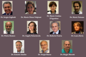
Basic Science | Volume 3 | Issue 1 | JRS Jun – June 2023 | Page 15-17 | Sergio Gigliotti , Maria Chiara Vulpiani Maria Cristina D’Agostino , Mario Vetrano, Pietro Romeo , Angela Notarnicola , Roberto Frairia , Laura Berta , Ernesto Amelio, Sergio Russo , Biagio Moretti.
DOI: 10.13107/jrs.2023.v03.i01.073
Author: Sergio Gigliotti [1], Maria Chiara Vulpiani [2], Maria Cristina D’Agostino [3], Mario Vetrano [2], Pietro Romeo [4], Angela Notarnicola [5], Roberto Frairia [6], Laura Berta [4], Ernesto Amelio [7], Sergio Russo [8], Biagio Moretti [5]
[1] Azienda Sanitaria Locale Napoli 1 Centro-Department of Orthopaedics, Campania, Italy,
[2] Department of Orthopaedics, University of Rome “La Sapienza”,Roma, Italy,
[3] Department of Orthopaedic Rehabilitation, Humanitas Research Hospital, Milan, Italy,
[4] Med and Sport 2000 Center, Turin, Italy,
[5] Department of Orthopaedics, University of Bari “Aldo Moro”, Bari, Italy,
[6] Department of Internal Medicine, University of Turin, Turin, Italy,
[7] Department of Orthopaedics, University of Verona, Italy,
[8] Department of Orthopaedics, University of Naples, Campania, Italy.
Address of Correspondence
Dr. Sergio Gigliotti,
ASL NA1Centro-Department of Orthopaedics, Campania, Italy.
Email: gigliotti_2004@libero.it
References:
- Mariotto S, Cavalieri E, Amelio E, Ciampa AR, de Prati AC, Marlinghaus E, et al. Extracorporeal shock waves: From lithotripsy to anti-inflammatory action by NO production. Nitric Oxide 2005;12:89-96.
- Berta L, Fazzari A, Ficco AM, Enrica PM, Catalano MG, Frairia R. Extracorporeal shock waves enhance normal fibroblast proliferation in vitro and activate mRNA expression for TGF-beta1 and for collagen Types I and III. Acta Orthop 2009;80:612-7.
- Catalano MG, Marano F, Rinella L, de Girolamo L, Bosco O, Fortunati N, et al. Extracorporeal shockwaves (ESWs) enhance the osteogenic medium-induced differentiation of adipose-derived stem cells into osteoblast-like cells. J Tissue Eng Regen Med 2017;11:390-9.
- Simplicio CL, Purita J, Murrell W, Santos GS, Dos Santos RG, Lana JF. Extracorporeal shock wave therapy mechanisms in musculoskeletal regenerative medicine. J Clin Orthop Trauma 2020; 11 Suppl 3:S309-18.
- Rinella L, Marano F, Berta L, Bosco O, Fraccalvieri M, Fortunati N, et al. Extracorporeal shock waves modulate myofibroblast differentiation of adipose-derived stem cells. Wound Repair Regen 2016;24:275-86.
- Muzio G, Vernè E, Canuto RA, Martinasso G, Saracino S, Baino F, et al. Shock waves induce activity of human osteoblast-like cells in bioactive scaffolds. J Trauma 2010;68:1439-44.
- De Girolamo L, Stanco D, Galliera E, Viganò M, Lovati AB, Marazzi MG, et al. Soft-focused extracorporeal shock waves increase the expression of tendon-specific markers and the release of anti-inflammatory cytokines in an adherent culture model of primary human tendon cells. Ultrasound Med Biol 2014;40:1204-15.
- Sansone V, D’ Agostino MC, Bonora C, Sizzano F, De Girolamo L, Romeo P. Early angiogenic response to shock waves in a three-dimensional model of human microvascular endothelial cell culture (HMEC-1). J Biol Regul Homeost Agents 2012;26:29-37.
- Vetrano M, D’Alessandro F, Torrisi MR, Ferretti A, Vulpiani MC, Visco V. Extracorporeal shock wave therapy promotes cell proliferation and collagen synthesis of primary cultured human tenocytes. Knee Surg Sports Traumatol Arthrosc 2011;19:2159-68.
- Leone L, Vetrano M, Ranieri D, Raffa S, Vulpiani MC, Ferretti A, et al. Extracorporeal Shock Wave Treatment (ESWT) improves in vitro functional activities of ruptured human tendon-derived tenocytes. PLoS One 2012;7:e49759.
- Leone L, Raffa S, Vetrano M, Ranieri D, Malisan F, Scrofani C, et al. Extracorporeal Shock Wave Treatment (ESWT) enhances the in vitro-induced differentiation of human tendon-derived stem/progenitor cells (hTSPCs) Oncotarget 2016;7:6410-23.
- Vetrano M, Ranieri D, Nanni M, Pavan A, Malisan F, Vulpiani MC, et al. Hyaluronic Acid (HA), Platelet-Rich Plasm and Extracorporeal Shock Wave Therapy (ESWT) promote human chondrocyte regeneration in vitro and ESWT-mediated increase of CD44 expression enhances their susceptibility to HA treatment. PLoS One 2019;14:e0218740.
- Moretti B, Iannone F, Notarnicola A, Lapadula G, Moretti L, Patella V, et al. Extracorporeal shock waves down-regulate the expression of interleukin-10 and tumor necrosis factor-alpha in osteoarthritic chondrocytes. BMC Musculoskelet Disord 2008;9:16.
- Tamma R, dell’Endice S, Notarnicola A, Moretti L, Patella S, Patella V, et al. Extracorporeal shock waves stimulate osteoblast activities. Ultrasound Med Biol 2009;35:2093-100.
- Iannone F, Moretti B, Notarnicola A, Moretti L, Patella S, Patella V, et al. Extracorporeal shock waves increase interleukin-10 expression by human osteoarthritic and healthy osteoblasts in vitro. Clin Exp Rheumatol 2009;27:794-9.
- Notarnicola A, Tamma R, Moretti L, Fiore A, Vicenti G, Zallone A, et al. Effects of radial shock waves therapy on osteoblasts activities Musculoskelet Surg 2012;96:183-9.
| How to Cite this article: Gigliotti S, Vulpiani MC, D’Agostino MC, Vetrano M, Romeo P, Notarnicola A, Frairia R, Berta L, Amelio E, Russo S, Moretti B. How shock waves works on musculoskeletal tissues? The contribution of Italian school. | Journal of Regenerative Science | Jan – June 2023; 3(1): 15-17. |
[Full Text HTML] [Full Text PDF]
In the Garden of Extracorporeal Shock Wave Therapy, Not Everything is Roses
Review Article | Volume 3 | Issue 1 | JRS Jun – June 2023 | Page 18-21 | Achim M. Loske
DOI: 10.13107/jrs.2023.v03.i01.075
Author: Achim M. Loske
[1] Centro de Física Aplicada y Tecnología Avanzada, Universidad Nacional Autónoma de México, Juriquilla, Querétaro, México.
Address of Correspondence
Dr. Achim M Loske,
Centro de Física Aplicada y Tecnología Avanzada, Universidad Nacional Autónoma de México,
Blvd. Juriquilla 3001, Querétaro, Qro., 76230 México.
E-mail: loske@fata.unam.mx
Abstract
The popularity of extracorporeal shock wave therapy to treat a large variety of medical conditions is indisputable. Despite this, sometimes poor results are obtained, and mild to severe complications have been reported. In most cases, wrong information and lack of training are responsible for this. The objective of this article is to explain the potential danger of using shock waves and radial pressure waves, as well as the reasons why, from the point of view of physics, sometimes the outcome is not as expected.
Keywords: Extracorporeal shock wave therapy, Shock waves, Radial pressure waves, Interaction with matter, Acoustic cavitation.
References:
- International Society for Medical Shockwave Therapy. Introduction and Prerequisites and Minimal Standards of Performing ESWT. Available from: https://www.shockwavetherapy.org/about-eswt/indications
- Novak KF. Physics: F-SW and R-SW. Basic information on focused and radial shock wave physics. In: Lohrer H, Gerdesmeyer L, editors. Multisdisciplinary Medical Applications. Heilbronn, Germany: Level 10 Buchverlag Daniela Bamberg; 2014.
- Philipp A, Delius M, Scheffczyk C, Vogel A, Lauterborn W. Interaction of lithotripter-generated shock waves with air bubbles. J Acoust Soc Am 1993;93:2496-509.
- Coralic V. Simulation of Shock-induced Bubble Collapse with Application to Vascular Injury in Shockwave Lithotripsy. Dissertation. Pasadena, CA, USA: California Institute of Technology; 2014.
- Ueberle F, Jamshidi Rad A. Ballistic pain therapy devices: Measurement of pressure pulse parameters. Biomed Eng/Biomed Tech 2012;57:700-3.
| How to Cite this article: Loske AM. In the Garden of Extracorporeal Shock Wave Therapy, Not Everything is Roses. | Journal of Regenerative Science | Jan – June 2023; 3(1): 18-21. |
[Full Text HTML] [Full Text PDF]
Extracorporeal Shockwave Therapy in Greater Trochanteric Pain Syndrome
Review Article | Volume 3 | Issue 1 | JRS Jun – June 2023 | Page 22-25 | Oyama Arruda Frei Caneca Junior , Ibrahim Afranio Willi Liu .
DOI: 10.13107/jrs.2023.v03.i01.077
Author: Oyama Arruda Frei Caneca Junior [1], Ibrahim Afranio Willi Liu [2]
[1] Director of SMBTOC, Orthopedic Surgeon at GOT Recife (Orthopedics and Traumatology Group), Brazil,
[2] Director of the Brazilian Medical Society for Shockwave Treatment-SMBTOC and Brazilian Orthopedic Society-SBOT Pain Committee.
Address of Correspondence
Dr. Oyama Arruda Frei Caneca Junior,
Director of SMBTOC, Orthopedic Physician at GOT Recife (Orthopedics and Traumatology Group), Brazil.
E-mail: oyama.arruda@gmail.com
Abstract
Peritrochanteric hip pain or great trochanter pain syndrome (GTPS) is a frequent complaint in offices and is the most common cause of pain and tenderness affecting the lateral part of the hip. Traditional conservative treatment of GTPS includes the use of anti-inflammatory drugs, physical therapy, and changing activities of daily living. In resistant cases, shockwave treatment presents satisfactory results considered good and excellent in 70 to 80% of GTPS cases treated by this technique, reducing the need for other treatments and the use of medications for long eriods. The treatment of GTPS with shock waves can be performed with focal waves or radial pressure waves, with the application of 3 initial sessions with an interval of one week, frequencies between 4 and 6 Hz, with 500 initial pulses in the region and at least 2000 pulses using energy between medium and high intensity at the point of greatest sensitivity on palpation, It is also important to treat the trigger points of the hip region. Due to the anatomical characteristics of a deep joint and the frequent presence of an associated myofascial pain, hip pathologies are a good option for the concomitant use of focal and radial pressure waves.
Keywords: Greater trochanteric pain syndrome; Trochanteric bursitis; hip pain; lateral hip pain; Shock waves; Radial pressure waves.
References:
- Long SS, Surrey DE, Nazarian LN. Sonography of greater trochanteric pain syndrome and the rarity of primary bursitis. AJR Am J Roentgenol. 2013 Nov;201(5):1083-6.
- Williams BS, Cohen SP. Greater trochanteric pain syndrome: a review of anatomy, diagnosis and treatment. Anesth Analg. 2009 May;108(5):1662-70.
- Pianka MA, Serino J, DeFroda SF, Bodendorfer BM. Greater trochanteric pain syndrome: Evaluation and management of a wide spectrum of pathology. SAGE Open Med. 2021 Jun 3;9:20503121211022582. doi: 10.1177/20503121211022582.
- Devin, C. J., McCullough, K. A., Morris, B. J., Yates, A. J., & Kang, J. D. (2012). Hip-spine Syndrome. Journal of the American Academy of Orthopaedic Surgeons, 20(7), 434–442.
- Donnelly JM. Travell, Simons & Simons’ myofascial pain and dysfunction: the trigger point manual. 3. ed. Philadelphia: Wolters Kluwer Health; 2019
- Gleitz, M., and K. Hornig. “Trigger Points–Diagnosis and treatment concepts with special reference to extracorporeal shockwaves.” Der Orthopäde41 (2012): 113-125.
- Alves FRV, Kruel AVS. Tratamento por ondas de choque nas patologias do quadril. In: Tratado de Ondas de Choque/ Soc Med Bras Trat Ondas de Choque. Ed. Alef, 2022.
- Wang Y, Wang K, Qin Y, Wang S, Tan B, Jia L, Jia G, Niu L. The effect of corticosteroid injection in the treatment of greater trochanter pain syndrome: a systematic review and meta-analysis of randomized controlled trials. J Orthop Surg Res. 2022 May 21;17(1):283. doi: 10.1186/s13018-022-03175-5
- Moya D, Ramón S, Schaden W, Wang CJ, Guiloff L, Cheng JH. The role of extracorporeal shockwave treatment in musculoskeletal disorders. J Bone Joint Surg Am. 2018 Feb 7;100(3):251-263. doi: 10.2106/JBJS.17.00661. PMID: 29406349.
- Furia JP, Rompe JD, Maffulli N. Low-energy extracorporeal shock wave therapy as a treatment for greater trochanteric pain syndrome. Am J Sports Med. 2009 Sep;37(9):1806-13. doi: 10.1177/0363546509333014.
- Ramon S, Russo S, Santoboni F, Lucenteforte G, Di Luise C, de Unzurrunzaga R, Vetrano M, Albano M, Baldini R, Cugat R, Stella G, Balato G, Seijas R, Nusca SM, Servodidio V, Vulpiani MC. Focused shockwave treatment for greater trochanteric pain syndrome: a multicenter, randomized, controlled clinical trial. J Bone Joint Surg Am. 2020 Aug 5;102(15):1305-1311. doi: 10.2106/JBJS.20.00093.
- Carlisi E, Cecini M, Di Natali G, Manzoni F, Tinelli C, Lisi C. Focused extracorporeal shock wave therapy for greater trochanteric pain syndrome with gluteal tendinopathy: a randomized controlled trial. Clin Rehabil. 2019 Apr;33(4):670-680. doi: 10.1177/0269215518819255.
- Schroeder AN, Tenforde AS, Jelsing EJ. Extracorporeal shockwave therapy in the management of sports medicine injuries. Curr Sports Med Rep. 2021 Jun 1;20(6):298-305. doi: 10.1249/JSR.0000000000000851.
- Mitchkash M, Robinson D, Tenforde AS. Efficacy of extracorporeal pulse-activated therapy in the management of lower-extremity running-related injuries: findings from a large case cohort. J Foot Ankle Surg. 2020 Jul-Aug;59(4):795-800. doi: 10.1053/j.jfas.2020.02.008.
- Congresso Brasileiro de Tratamento por Ondas de Choque, 3º, 2018, São Paulo. Consensos no Tratamento da Síndrome Trocantérica. III Brazilian Congress of Shockwave Treatment (SMBTOC).
- Cowan RM, Semciw AI, Pizzari T, Cook J, Rixon MK, Gupta G, Plass LM, Ganderton CL. Muscle size and quality of the gluteal muscles and tensor fasciae latae in women with greater trochanteric pain syndrome. Clin Anat. 2020 Oct;33(7):1082-1090. doi: 10.1002/ca.23510. Epub 2019 Nov 24.

| How to Cite this article: Junior OAFC, Liu IAW | Extracorporeal Shockwave Therapy in Greater Trochanteric Pain Syndrome. | Journal of Regenerative Science | Jan – June 2023; 3(1): 22-25. |
[Full Text HTML] [Full Text PDF]
Achilles Tendinopathy, Pathophysiology, Diagnosis, and Management with Shockwave Therapy
Review Article | Volume 3 | Issue 1 | JRS Jun – June 2023 | Page 26-31 | Paul German Terán , Estefania Anabel Lozada , Alvaro Santiago LeMarie.
DOI: 10.13107/jrs.2023.v03.i01.79
Author: Paul German Terán [1], Estefania Anabel Lozada [1], Alvaro Santiago LeMarie [2]
[1] Department of Traumatology and Orthopedics, Orthopedic Specialties Center, Quito, Ecuador,
[2] Anatomy Professor, Faculty of Medicine, International University of Ecuador.
Address of Correspondence
Dr. Paul German Terán,
Department of Traumatology and Orthopedics, Orthopedic Specialties Center, Quito, Ecuador.
E-mail: paulteranmd@gmail.com
Abstract
The Achilles tendon is a strong structure that is frequently injured in runners and jumpers, but it can also be present in patients who do not engage in any sports. This clinical syndrome is characterized by pain, structural changes, and impairment of physical function. Achilles tendinopathy is extensively studied because it can be devastating, with slow and prolonged recovery that can take a year or more, and a high risk of re-injury. This condition is classified into insertional and non-insertional Achilles tendinopathy, depending on the affected region of the tendon. Intrinsic and extrinsic factors contribute to the intratendinous changes in vascularization and elevated pain neurotransmitters. The diagnosis is primarily based on the patient’s clinical history, and imaging techniques such as ultrasound, including sonoelastography, and magnetic resonance imaging can be useful in identifying the nature, location, and extent of the lesion. Treatment options for Achilles tendinopathy include rehabilitation, image-guided injections, shockwave therapy, ultrasound therapy, percutaneous intratissue electrolysis, orthotics, medications, and surgery. Among these options, shockwave therapy may provide the best tolerance, pain relief, and functional recovery.
Keywords: Achilles tendon, tendinopathy, insertional, non-insertional, extracorporeal shock waves.
References:
- Weinfeld SB. Achilles tendon disorders. Vol. 98, Medical Clinics of North America. 2014. p. 331–8.
- Silbernagel KG, Hanlon S, Sprague A. Current clinical concepts: Conservative management of achilles tendinopathy. J Athl Train. 2020 May 1;55(5).
- Li HY, Hua YH. Achilles Tendinopathy: Current Concepts about the Basic Science and Clinical Treatments. Vol. 2016, BioMed Research International. Hindawi Limited; 2016.
- Lysholm J, Wiklander J. Injuries in runners.
- Li HY, Hua YH. Achilles Tendinopathy: Current Concepts about the Basic Science and Clinical Treatments. Vol. 2016, BioMed Research International. Hindawi Limited; 2016.
- Li HY, Hua YH. Achilles Tendinopathy: Current Concepts about the Basic Science and Clinical Treatments. Vol. 2016, BioMed Research International. Hindawi Limited; 2016.
- Maffulli N, Longo UG, Kadakia A, Spiezia F. Achilles tendinopathy. Vol. 26, Foot and Ankle Surgery. Elsevier Ltd; 2020. p. 240–9.
- Grävare Silbernagel K, Malliaras P, de Vos RJ, Hanlon S, Molenaar M, Alfredson H, et al. ICON 2020—International Scientific Tendinopathy Symposium Consensus: A Systematic Review of Outcome Measures Reported in Clinical Trials of Achilles Tendinopathy. Vol. 52, Sports Medicine. Springer Science and Business Media Deutschland GmbH; 2022. p. 613–41.
- Färnqvist K, Pearson S, Malliaras P. Adaptation of Tendon Structure and Function in Tendinopathy With Exercise and Its Relationship to Clinical Outcome. Vol. 29, Journal of Sport Rehabilitation. Human Kinetics Publishers Inc.; 2020. p. 107–15.
- Del Buono A, Chan O, Maffulli N. Achilles tendon: Functional anatomy and novel emerging models of imaging classification. Int Orthop. 2013 Apr;37(4):715–21.
- Thomopoulos S, Parks WC, Rifkin DB, Derwin KA. Mechanisms of tendon injury and repair. In: Journal of Orthopaedic Research. John Wiley and Sons Inc.; 2015. p. 832–9.
- Li HY, Yasui Y, Han SH, Miyamoto W, Hua YH. Achilles Tendinopathy: From the Basic Science to the Clinic. Vol. 2017, BioMed Research International. Hindawi Limited; 2017.
- Magnan B, Bondi M, Pierantoni S, Samaila E. The pathogenesis of Achilles tendinopathy: A systematic review. Vol. 20, Foot and Ankle Surgery. Elsevier Ltd; 2014. p. 154–9.
- Van Der Vlist AC, Breda SJ, Oei EHG, Verhaar JAN, De Vos RJ. Clinical risk factors for Achilles tendinopathy: A systematic review. Vol. 53, British Journal of Sports Medicine. BMJ Publishing Group; 2019. p. 1352–61.
- Ackermann PW, Renström P. Tendinopathy in Sport. Sports Health. 2012 May;4(3):193–201.
- Rai V, Dietz NE, Dilisio MF, Radwan MM, Agrawal DK. Vitamin D attenuates inflammation, fatty infiltration, and cartilage loss in the knee of hyperlipidemic microswine. Arthritis Res Ther. 2016 Sep 13;18(1).
- Klein EE, Weil L, Weil LS, Fleischer AE. Body Mass Index and Achilles Tendonitis: A 10-Year Retrospective Analysis. Foot Ankle Spec. 2013 Aug;6(4):276–82.
- Masci L, Spang C, Van Schie HTM, Alfredson H. How to diagnose plantaris tendon involvement in midportion Achilles tendinopathy – Clinical and imaging findings. BMC Musculoskelet Disord. 2016 Feb 24;17(1).
- Geyer M. Achillodynie. Orthopade. 2005 Jul;34(7):677–81.
- Khaliq Y, Zhanel GG. Fluoroquinolone-Associated Tendinopathy: A Critical Review of the Literature.
- Singh D. Cholesterol level in non-insertional Achilles tendonopathy. Foot. 2015 Dec 1;25(4):228–31.
- Luck MD, Gordon AG, Blebea JS, Dalinka MK. High association between accessory soleus muscle and Achilles tendonopathy. Skeletal Radiol. 2008 Dec;37(12):1129–33.
- Vallone G, Vittorio T. Complete Achilles tendon rupture after local infiltration of corticosteroids in the treatment of deep retrocalcaneal bursitis. J Ultrasound. 2014;17(2):165–7.
- Abate M, Salini V, Schiavone C. Achilles tendinopathy in elderly subjects with type II diabetes: the role of sport activities. Aging Clin Exp Res. 2016 Apr 1;28(2):355–8.
- Magnan B, Bondi M, Pierantoni S, Samaila E. The pathogenesis of Achilles tendinopathy: A systematic review. Vol. 20, Foot and Ankle Surgery. Elsevier Ltd; 2014. p. 154–9.
- Marques PP, Vieira CP, de Oliveira LP, Pimentel ER, Guerra FDR. Chronical treatment with sildenafil causes Achilles tendinopathy in rats. Life Sci. 2018 Nov 1;212:87–92.
- Docking SI, Rosengarten SD, Daffy J, Cook J. Structural integrity is decreased in both Achilles tendons in people with unilateral Achilles tendinopathy. J Sci Med Sport. 2015;18(4):383–7.
- Chimenti RL, Cychosz CC, Hall MM, Phisitkul P. Current Concepts Review Update: Insertional Achilles Tendinopathy. Foot Ankle Int. 2017 Oct 1;38(10):1160–9.
- Gaston TE, Daniel JN. Achilles insertional tendinopathy – Is there a gold standard? Vol. 9, Archives of Bone and Joint Surgery. Mashhad University of Medical Sciences; 2021. p. 5–8.
- Grambart ST, Lechner J, Wentz J. Differentiating Achilles Insertional Calcific Tendinosis and Haglund’s Deformity. Vol. 38, Clinics in Podiatric Medicine and Surgery. W.B. Saunders; 2021. p. 165–81.
- Jarin I, Bäcker HC, Vosseller JT. Meta-analysis of Noninsertional Achilles Tendinopathy. Vol. 41, Foot and Ankle International. SAGE Publications Inc.; 2020. p. 744–54.
- Pearce CJ, Tan A. Non-insertional Achilles tendinopathy. EFORT Open Rev. 2016 Nov 1;1(11):383–90.
- De Vos RJ, Van Der Vlist AC, Winters M, Van Der Giesen F, Weir A. Diagnosing Achilles tendinopathy is like delicious spaghetti carbonara: It is all about key ingredients, but not all chefs use the same recipe. Vol. 55, British Journal of Sports Medicine. BMJ Publishing Group; 2021. p. 247–8.
- Pierre-Jerome C, Moncayo V, Terk MR. MRI of the Achilles tendon: A comprehensive review of the anatomy, biomechanics, and imaging of overuse tendinopathies. Vol. 51, Acta Radiologica. 2010. p. 438–54.
- Lopez RGL, Jung HG. Achilles tendinosis: Treatment options. CiOS Clinics in Orthopedic Surgery. 2015 Mar 1;7(1):1–7.
- Knobloch K. Drug-induced tendon disorders. In: Advances in Experimental Medicine and Biology. Springer New York LLC; 2016. p. 229–38.
- Bolon B. Mini-Review: Toxic Tendinopathy. Vol. 45, Toxicologic Pathology. SAGE Publications Inc.; 2017. p. 834–7.
- Taneja AK, Santos DCB. Steroid-induced Kager’s fat pad atrophy. Vol. 43, Skeletal Radiology. Springer Verlag; 2014. p. 1161–4.
- Aicale R, Oliviero A, Maffulli N. Management of Achilles and patellar tendinopathy: What we know, what we can do. Vol. 13, Journal of Foot and Ankle Research. BioMed Central Ltd; 2020.
- Kyle Duchman, Devin Lemmex, Sunny Patel, Grant Garrigues, Jonathan Riboh. The Effect of Non-Steroidal Anti-Inflammatory Drugs on Tendon-to-Bone Healing: A Systematic Review with Subgroup Meta-Analysis. Iowa Orthopaedic Journal. 2019;39(1):107–19.
- Scott A, Bahr R. Neuropeptides in tendinopathy. 2014.
- Hijlkema A, Roozenboom C, Mensink M, Zwerver J. The impact of nutrition on tendon health and tendinopathy: a systematic review. Vol. 19, Journal of the International Society of Sports Nutrition. Taylor and Francis Ltd.; 2022. p. 474–504.
- Fusini F, Bisicchia S, Bottegoni C, Gigante A, Zanchini F, Busilacchi A. Nutraceutical supplement in the management of tendinopathies: a systematic review. 2016.
- Van Der Vlist AC, Winters M, Weir A, Ardern CL, Welton NJ, Caldwell DM, et al. Which treatment is most effective for patients with Achilles tendinopathy? A living systematic review with network meta-analysis of 29 randomised controlled trials. Vol. 55, British Journal of Sports Medicine. BMJ Publishing Group; 2021. p. 249–55.
- Balius R, Álvarez G, Baró F, Jiménez F, Pedret C, Costa E, et al. A 3-Arm Randomized Trial for Achilles Tendinopathy: Eccentric Training, Eccentric Training Plus a Dietary Supplement Containing Mucopolysaccharides, or Passive Stretching Plus a Dietary Supplement Containing Mucopolysaccharides. Curr Ther Res Clin Exp. 2016;78:1–7.
- Gatz M, Betsch M, Dirrichs T, Schrading S, Tingart M, Michalik R, et al. Eccentric and Isometric Exercises in Achilles Tendinopathy Evaluated by the VISA-A Score and Shear Wave Elastography. Sports Health. 2020 Jul 1;12(4):373–81.
- Färnqvist K, Pearson S, Malliaras P. Adaptation of Tendon Structure and Function in Tendinopathy With Exercise and Its Relationship to Clinical Outcome. Vol. 29, Journal of Sport Rehabilitation. Human Kinetics Publishers Inc.; 2020. p. 107–15.
- Tumilty S, Mani R, Baxter GD. Photobiomodulation and eccentric exercise for Achilles tendinopathy: a randomized controlled trial. Lasers Med Sci. 2016 Jan 1;31(1):127–35.
- Abat F, Diesel WJ, Gelber PE, Polidori F, Monllau JC, Sanchez-Ibañez JM. Effectiveness of the Intratissue Percutaneous Electrolysis (EPI®) technique and isoinertial eccentric exercise in the treatment of patellar tendinopathy at two years follow-up. Vol. 4, Ligaments and Tendons Journal. 2014.
- Fan Y, Feng Z, Cao J, Fu W. Efficacy of Extracorporeal Shock Wave Therapy for Achilles Tendinopathy: A Meta-analysis. Vol. 8, Orthopaedic Journal of Sports Medicine. SAGE Publications Ltd; 2020.
- Gerdesmeyer L, Mittermayr R, Fuerst M, Al Muderis M, Thiele R, Saxena A, et al. Current evidence of extracorporeal shock wave therapy in chronic Achilles tendinopathy. Vol. 24, International Journal of Surgery. Elsevier Ltd; 2015. p. 154–9.
- Benli MD, Tatari H, Balcı A, Peker A, Şimşek K, Yüksel O, et al. A comparison between the efficacy of eccentric exercise and extracorporeal shock wave therapy on tendon thickness, vascularity, and elasticity in Achilles tendinopathy: A randomized controlled trial. Turk J Phys Med Rehabil. 2022;68(3):372–80.
- Hasselbalch L. Ekstrakorporal shockbølgeterapi ved langvarig akillessenetendinopati.
- Zhang S, Li H, Yao W, Hua Y, Li Y. Therapeutic Response of Extracorporeal Shock Wave Therapy for Insertional Achilles Tendinopathy Between Sports-Active and Nonsports-Active Patients With 5-Year Follow-up. Orthop J Sports Med. 2020 Jan 1;8(1).
- Stania M, Juras G, Chmielewska D, Polak A, Kucio C, Król P, et al. Extracorporeal Shock Wave Therapy for Achilles Tendinopathy. 2019.
- Insuasti Abarca W, Llocclla Delgado S, Terán Vela P, Platero Portillo T, Martínez Asnalema D, Abarca García L. Patellar tendon rupture after radial pressure wave therapy – also known as radial shock wave therapy – for patellar tendinopathy: Report of two Cases. Revista de la Facultad de Medicina Humana. 2021 Mar 15;21(2):449–58.
- Terán PG, Insuasti WE, Martínez DM, Platero TM, Ramos SG, Llocclla S. Lesión del nervio cubital secundario a terapia de ondas de choque extracorpóreas radiales identificada con ultrasonografía de alta resolución: Reporte de caso. Revista de la Facultad de Medicina Humana. 2020 Mar 27;20(2):156–61.
- Moya D, Ramón S, Guiloff L, Terán P, Eid J, Serrano E. Poor results and complications in the use of focused shockwaves and radial pressure waves in musculoskeletal pathology. Vol. 56, Rehabilitacion. Ediciones Doyma, S.L.; 2022. p. 64–73.
- Stenson JF, Reb CW, Daniel JN, Saini SS, Albana MF. Predicting Failure of Nonoperative Treatment for Insertional Achilles Tendinosis. Foot Ankle Spec. 2018 Jun 1;11(3):252–5.

| How to Cite this article: Terán PG, Lozada EA, LeMarie AS. | Achilles Tendinopathy, Pathophysiology, Diagnosis, and Management with Shockwave Therapy. | Journal of Regenerative Science | Jan – June 2023; 3(1): 26-31. |
[Full Text HTML] [Full Text PDF]
Lateral Epicondylitis: General Concepts and Shock Wave Treatment Evidence
Review Article | Volume 3 | Issue 1 | JRS Jun – June 2023 | Page 32-34 | Ricardo Kobayashi.
DOI: 10.13107/jrs.2023.v03.i01.81
Author: Ricardo Kobayashi [1]
[1] Pain Center, University of S£o Paulo, S£o Paulo, Brazil.
Address of Correspondence
Dr. Ricardo Kobayashi, MD, Phd,
Pain Center, University of S£o Paulo, S£o Paulo, Brazil.
E-mail: institutokobayashi@gmail.com
Abstract
Introduction:Lateral epicondylitis (LE) is one of the most common tendinopathies of the upper extremity characterized by lateral elbow pain, seriously affecting patients’ daily life and work.
Pathophysiology: Anatomically, the common extensor insertion on the lateral epicondyle of the humerus, mostly the extensor carpi radial is brevis tendon insertion, undergoes microtearing associated with a chronic repair process, but hardly any inflammation. The pathoanatomy of overuse tendinopathy is non-inflammatory angiofibroblastic tendinosis. For this reason, the term ‘‘tendinitis’’ is avoided, and ‘‘tendinosis’’ is preferred.
Diagnosis: LE is primarily a clinical diagnosis. The natural history is a gradual onset of pain in the absence of defined trauma. The most
common findings on physical examination are tenderness at the lateral epicondyle of the distal humerus and weakness or pain with resisted wrist extension (the Thomsen test).
Treatment: Non-surgical options are the mainstream treatment for LE, a small proportion of patients eventually undergoes surgery, although surgery for LE is no more effective than non-surgical treatment, based on evidence. Non-operative treatments including rest, application of ice, administration of analgesic medications, orthopedic devices, ultrasound, transcutaneous electrical nerve stimulation, eccentric training, and extracorporeal shock wave therapy (ESWT).
Shockwave Treatment of LE: There are many therapeutic options for treating LE. The existing evidence does not clearly support the efficacy of any of the available treatment methods for this clinical condition. ESWT is not the exception, although it was approved by the U.S. Food and Drug Administration for treating this disease in 2002 and much of the current evidence supports its indication for LE..
Keywords: Lateral epicondylitis, Tennis elbow, Tendinopathy, Shock waves.
References:
- Wolf JM. Lateral epicondylitis. N Engl J Med 2023;388:2371-7.
- Marigi EM, Dancy M, Alexander A, Marigi IM, Clark J, Krych AJ, et al. Lateral epicondylitis: Critical analysis review of current nonoperative treatments. JBJS Rev 2023;11(2): doi: 10.2106/JBJS.RVW.22.00170
- Thiele S, Thiele R, Gerdesmeyer L. Lateral epicondylitis: This is still a main indication for extracorporeal shockwave therapy. Int J Surg 2015;24:165-70.
- Nirschl RP, Ashman ES. Elbow tendinopathy: Tennis elbow. Clin Sports Med 2003;22:813-36.
- Xu J, Chen M, Xue X, Zhou W, Luo X. Global research trends and hotspots in lateral epicondylitis during the past 30 Years: A bibliometric and visualization study. Med Sci Monit 2023;29:e939309.
- Yao G, Chen J, Duan Y, Chen X. Efficacy of extracorporeal shock wave therapy for lateral epicondylitis: A systematic review and meta-analysis. Biomed Res Int 2020;2020:2064781.
- van Leeuwen WF, Janssen SJ, Ring D, Chen N. Incidental magnetic resonance imaging signal changes in the extensor carpi radialis brevis origin are more common with age. J Shoulder Elbow Surg 2016;25:1175-81.
- van Kollenburg JA, Brouwer KM, Jupiter JB, Ring D. Magnetic resonance imaging signal abnormalities in enthesopathy of the extensor carpi radialis longus origin. J Hand Surg Am 2009;34:1094-8.
- Liu WC, Chen CT, Lu CC, Tsai YC, Liu YC, Hsu CW, et al. Extracorporeal shock wave therapy shows superiority over injections for pain relief and grip strength recovery in lateral epicondylitis: A systematic review and network meta-analysis. Arthroscopy 2022;38:2018-34.e12.
- Yoon SY, Kim YW, Shin IS, Moon HI, Lee SC. Does the type of extracorporeal shock therapy influence treatment effectiveness in lateral epicondylitis? A systematic review and meta-analysis. Clin Orthop Relat Res 2020;478:2324-39.
- Moya D, Ramón S, Schaden W, Wang CJ, Guiloff L, Cheng JH. The role of extracorporeal shockwave treatment in musculoskeletal disorders. J Bone Joint Surg Am 2018;100:251-63.
- Camargo LS, Kobayashi R. Case Report: Extracorporeal shock wave treatment in plantar fasciitis with an associated neuropathic component. How to optimize the result? J Regen Sci 2022;2:21-3.
- Haake M, Konig IR, Decker T, Riedel C, Buch M, Muller HH. Extracorporeal shock wave therapy in the treatment of lateral epicondylitis: A randomized multicenter trial. J Bone Joint Surg Am 2002;84:1982-91.
- Pettrone FA, McCall BR. Extracorporeal shock wave therapy without local anesthesia for chronic lateral epicondylitis. J Bone Joint Surg Am 2005;87:1297-304.
- Buchbinder R, Green SE, Youd JM, Assendelft WJ, Barnsley L, Smidt N. Shock wave therapy for lateral elbow pain. Cochrane Database Syst Rev 2005;4:CD003524.
- Moghtaderi A, Khosrawi S, Dehghan F. Extracorporeal shock wave therapy of gastroc-soleus trigger points in patients with plantar fasciitis: A randomized, placebo-controlled trial. Adv Biomed Res 2014;3:99.

| How to Cite this article: Kobayashi R. | Lateral Epicondylitis: General Concepts and Shock Wave Treatment Evidence. | Journal of Regenerative Science | Jan – June 2023; 3(1): 32-34. |