Posts

Analysis of therapeutic effect of high focused extracorporeal shock wave comprehensive therapy on femoral head bone marrow edema syndrome

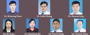
Original Article | Vol 3 | Issue 2 |  July-December 2023 | page: 35-40 | Ruimeng Duan, Leilei Zhang, Haonan Ling, Jie Guan, Huisheng Shi, Dawei Liang, Xiantao Chen

DOI: https://doi.org/10.13107/jrs.2023.v03.i02.99


Author: Ruimeng Duan [1], Leilei Zhang [1], Haonan Ling [1], Jie Guan [1], Huisheng Shi [1], Dawei Liang [1], Xiantao Chen [1]

[1] Department of Femoral Head Necrosis, Luoyang Orthopedic-Traumatological Hospital of Henan Province (Henan Provincial Orthopedic Hospital), Luoyang, Henan, China.

Address of Correspondence
Dr. Xiantao Chen,
Department of Femoral Head Necrosis, Luoyang Orthopedic-Traumatological Hospital of Henan Province (Henan Provincial Orthopedic Hospital), No. 82 Qiming South Road, Luoyang, Henan Province 471000, China.
E-mail: luoyangzhenggu@163.com


Abstract

Purpose: This study explored the clinical therapeutic effect of high-focused extracorporeal shock wave therapy (HF-ESWT) combined with exercise rehabilitation and drug therapy on femoral head bone marrow edema syndrome (BMES).
Materials and Methods: This study systematically reviewed and analyzed the clinical data of 43 patients with femoral head bone marrow edema who were treated in our hospital from January 2017 to June 2022. Twenty-three patients received HF-ESWT comprehensive treatment. Twenty patients received general treatment including medication and exercise rehabilitation treatment. The treatment methods for Group B patients were the same as Group A, except for not receiving shock wave therapy. Changes in visual analog scale (VAS), Harris score of the hip, and the edema area of region of interest area (ROIA) on hip magnetic resonance imaging (MRI) were analyzed before and after treatment.
Results: Our research found that patients receiving HF-ESWT had significantly reduced VAS compared with general treatment at 1, 2, and 3 months (P < 0.05). We found that HF-ESWT comprehensive treatment had significantly improved hip Harris score compared with general treatment at 2 and 3 months (P < 0.05). HF-ESWT comprehensive treatment had significantly reduced edema area of ROIA on hip MRI compared with general treatment at 1, 2, and 3 months (P < 0.05). In addition, the healing rate was significantly higher in the HF-ESWT
comprehensive treatment group compared with general treatment group (P < 0.05). One of the patients in the group treated with shockwaves developed hip pain that worsened after treatment, three patients developed local skin ecchymosis, and the other patients had no adverse events.
Conclusion: HF-ESWT comprehensive treatment significantly reduced hip pain symptoms, quickly shortened the time for femoral head edema to dissipate, and significantly improved hip function for affected limbs with bone marrow edema syndrome. HF-ESWT comprehensive treatment may be an effective therapeutic strategy for HF-BMES.
Keywords: Extracorporeal shock wave therapy, Bone marrow edema syndrome, Traditional Chinese medicine, Osteonecrosis of the femoral head


References:

1. Miyanishi K, Yamamoto T, Nakashima Y, Shuto T, Jingushi S, Noguchi Y, et al. Subchondral changes in transient osteoporosis of the hip. Skeletal Radiol 2001;30:255-61.
2. Guerra JJ, Steinberg ME. Distinguishing transient osteoporosis from avascular necrosis of the hip. J Bone joint Surg Am 1995;77:616-24.
3. Mirghasemi SA, Trepman E, Sadeghi MS, Rahimi N, Rashidinia S. Bone marrow edema syndrome in the foot and ankle. Foot Ankle Int 2016;37:1364-73.
4. Hofmann S. The painful bone marrow edema syndrome of the hip joint. Wien Klin Wochenschr 2005;117:111-20.
5. Hayes CW, Conway WF, Daniel WW. MR imaging of bone marrow edema pattern: transient osteoporosis, transient bone marrow edema syndrome, or osteonecrosis. Radiographics 1993;13:1001-11; discussion 1012.
6. Cui Q, Jo WL, Koo KH, Cheng EY, Drescher W, Goodman SB, et al. ARCO consensus on the pathogenesis of non-traumatic osteonecrosis of the femoral head. J Korean Med Sci 2021;36:e65.
7. Zhao D, Zhang F, Wang B, Liu B, Li L, Kim SY, et al. Guidelines for clinical diagnosis and treatment of osteonecrosis of the femoral head in adults (2019 version). J Orthop Transl 2020;21:100-10.
8. Eidmann A, Eisert M, Rudert M, Stratos I. Influence of vitamin D and C on bone marrow edema syndrome-a scoping review of the literature. J Clin Med 2022;11:6820.
9. Sansone V, Ravier D, Pascale V, Applefield R, Del Fabbro M, Martinelli N. Extracorporeal shockwave therapy in the treatment of nonunion in long bones: A systematic review and meta-analysis. J Clin Med 2022;11:1977.
10. Simon MJ, Barvencik F, Luttke M, Amling M, Mueller-Wohlfahrt HW, Ueblacker P. Intravenous bisphosphonates and vitamin D in the treatment of bone marrow oedema in professional athletes. Injury 2014;45:981-7.
11. Gao F, Sun W, Li Z, Guo W, Wang W, Cheng L, et al. Extracorporeal shock wave therapy in the treatment of primary bone marrow edema syndrome of the knee: A prospective randomised controlled study. BMC Musculoskelet Disord 2015;16:379.
12. Meng K, Liu Y, Ruan L, Chen L, Chen Y, Liang Y. Suppression of apoptosis in osteocytes, the potential way of natural medicine in the treatment of osteonecrosis of the femoral head. Biomed Pharmacother 2023;162:114403.
13. Qian D, Zhou H, Fan P, Yu T, Patel A, O’Brien M, et al. A traditional Chinese medicine plant extract prevents alcohol-induced osteopenia. Front Pharmacol 2021;12:754088.
14. Qi ZX, Chen L. Effect of Chinese drugs for promoting blood circulation and eliminating blood stasis on vascular endothelial growth factor expression in rabbits with glucocorticoid-induced ischemic necrosis of femoral head. J Tradit Chin Med 2009;29:137-40.
15. Yong EL, Logan S. Menopausal osteoporosis: Screening, prevention and treatment. Singapore Med J 2021;62:159-66.
16. Hofmann S, Engel A, Neuhold A, Leder K, Kramer J, Plenk H Jr. Bone-marrow oedema syndrome and transient osteoporosis of the hip. An MRI-controlled study of treatment by core decompression. J Bone Joint Surg Br 1993;75:210-6.
17. Schweitzer ME, White LM. Does altered biomechanics cause marrow edema? Radiology 1996;198:851-3.
18. Woertler K, Neumann J. Atraumatic bone marrow edema involving the epiphyses. Sem Musculoskelet Radiol 2023;27:45-53.
19. Plenk H Jr., Hofmann S, Eschberger J, Gstettner M, Kramer J, Schneider W, et al. Histomorphology and bone morphometry of the bone marrow edema syndrome of the hip. Clin Orthop Relat Res 1997;334:73-84.
20. Oehler N, Mussawy H, Schmidt T, Rolvien T, Barvencik F. Identification of vitamin D and other bone metabolism parameters as risk factors for primary bone marrow oedema syndrome. BMC Musculoskelet Disord 2018;19:451.
21. Petek D, Hannouche D, Suva D. Osteonecrosis of the femoral head: pathophysiology and current concepts of treatment. EFORT Open Rev 2019;4:85-97.
22. Miranian D, Lanham N, Stensby DJ, Diduch D. Progression and treatment of bilateral knee bone marrow edema syndrome. JBJS Case Connect 2015;5:e391-7.
23. Daly RM, Dalla Via J, Duckham RL, Fraser SF, Helge EW. Exercise for the prevention of osteoporosis in postmenopausal women: An evidence-based guide to the optimal prescription. Braz J Phys Ther 2019;23:170-80.
24. Vasiliadis AV, Zidrou C, Charitoudis G, Beletsiotis A. Single-dose therapy of zoledronic acid for the treatment of primary bone marrow edema syndrome. Cureus 2021;13:e13977.
25. Zippelius T, Strube P, Rohe S, Schlattmann P, Dobrindt O, Caffard T, et al. The use of iloprost in the treatment of bone marrow edema syndrome of the proximal femur: A review and meta-analysis. J Pers Med 2022;12:1757.
26. Gao F, Sun W, Li Z, Guo W, Kush N, Ozaki K. Intractable bone marrow edema syndrome of the hip. Orthopedics 2015;38:e263-70.
27. Mei J, Pang L, Jiang Z. The effect of extracorporeal shock wave on osteonecrosis of femoral head: A systematic review and meta-analysis. Phys Sportsmed 2022;50:280-8.
28. Yang X, Shi L, Zhang T, Gao F, Sun W, Wang P, et al. High-energy focused extracorporeal shock wave prevents the occurrence of glucocorticoid-induced osteonecrosis of the femoral head: A prospective randomized controlled trial. J Orthop Translat 2022;36:145-51.
29. Xie K, Mao Y, Qu X, Dai K, Jia Q, Zhu Z, et al. High-energy extracorporeal shock wave therapy for nontraumatic osteonecrosis of the femoral head. J Orthop Surg Res 2018;13:25.
30. Li B, Wang R, Huang X, Ou Y, Jia Z, Lin S, et al. Extracorporeal shock wave therapy promotes osteogenic differentiation in a rabbit osteoporosis model. Front Endocrinol 2021;12:627718.
31. Ma HZ, Zeng BF, Li XL. Upregulation of VEGF in subchondral bone of necrotic femoral heads in rabbits with use of extracorporeal shock waves. Calcifi Tissue Int 2007;81:124-31.
32. Huang HM, Li XL, Tu SQ, Chen XF, Lu CC, Jiang LH. Effects of roughly focused extracorporeal shock waves therapy on the expressions of bone morphogenetic protein-2 and osteoprotegerin in osteoporotic fracture in rats. Chin Med J (Engl) 2016;129:2567-75.
33. Yu T, Zhang Z, Xie L, Ke X, Liu Y. The influence of traditional Chinese medicine constitutions on the potential repair capacity after osteonecrosis of the femoral head. Complement Ther Med 2016;29:89-93.
34. Weng B, Chen C. Effects of bisphosphonate on osteocyte proliferation and bone formation in patients with diabetic osteoporosis. Comput Math Methods Med 2022;2022:2368564.
35. Liu P, Tu J, Wang W, Li Z, Li Y, Yu X, et al. Effects of mechanical stress stimulation on function and expression mechanism of osteoblasts. Front Bioeng Biotechnol 2022;10:830722.
36. Iolascon G, Resmini G, Tarantino U. Mechanobiology of bone. Aging Clin Exp Res 2013;25:S3-7.
37. Benton MJ, White A. Osteoporosis: Recommendations for resistance exercise and supplementation with calcium and vitamin D to promote bone health. J Community Health Nurs 2006;23:201-11.

How to Cite this article: Duan R, Zhang L, Ling H, Guan J, Shi H, Liang D, Chen X | Analysis of therapeutic effect of high focused extracorporeal shock wave comprehensive therapy on femoral head bone marrow edema syndrome | Journal of Regenerative Science | Jul-Dec 2023; 3(2): 35-40.

[Full Text HTML] [Full Text PDF] 


A novel treatment method for ankylosing spondylitis combined with sacroiliac joint bone marrow edema

Original Article | Vol 3 | Issue 2 |  July-December 2023 | page: 41-46 | Leilei Zhang, Xuanye Zhu, Haonan Ling, Wanyi Zhang, Ying Zhang, Youwen Liu, Xiantao Chen

DOI: https://doi.org/10.13107/jrs.2023.v03.i02.101


Author: Leilei Zhang [1], Xuanye Zhu [2], Haonan Ling [1], Wanyi Zhang [1], Ying Zhang [1], Youwen Liu [1], Xiantao Chen [1]

[1] Center of hip Surgery, Henan Luoyang Orthopedic-Traumatological Hospital, Orthopedic Hospital of Henan Province, Luoyang, China.
[2] Henan University of Traditional Chinese Medicine, zhengzhou, China.

Address of Correspondence
Dr. Center of Hip Surgery, Henan Luoyang Orthopedic-Traumatological Hospital, Orthopedic Hospital of Henan Province, No. 82, South Qiming Road, 471002, Luoyang, China.
E-mail: luoyangzhenggu@139.com


Abstract

Objective: To investigate whether high-energy extracorporeal shock wave therapy (ESWT) combined with conventional oral medicine as a potential novel therapeutic approach for the treatment of ankylosing spondylitis (AS)combined with sacroiliac joint bone marrow edema.
Materials & Methods: 40 patients were divided into two groups and were treated with or without ESWT in combination with conventional oral medicine. A visual analog scale (VAS) score of spinal pain, as well as indicators of spinal mobility, Bath Ankylosing Spondylitis Disease Activity Index (BASDAI) and Bath Ankylosing Spondylitis Functional Index (BASFI) scores, inflammatory index (C-reactive protein, blood cell sedimentation rate), and other indicators were compared between the two groups. The Spondyloarthritis Research Consortium of Canada (SPARCC) scoring system was used to evaluate pain and structural damage in the sacroiliac joint.
Results: (1) After one month of treatment (T1), VAS, BASDAI, BASFI, and SPARCC scores were lower in both groups than at the start of treatment (T0) (P < 0.05), with greater decreases observed in the treatment group (P < 0.05). (2) Also, at T1, indicators of spinal mobility for the two groups were improved (P < 0.05). (3) ESR and C-reactive protein levels for the two groups decreased significantly at T1 versus T0 (P < 0.05).
Conclusion: ESWT combined with oral medication can significantly relieve pain and improve clinical functional symptoms for patients with AS. It can also reduce sacroiliac joint bone marrow edema and control the inflammatory reaction in the sacroiliac joint, which represents a novel, effective, reliable, and safe clinical treatment therapeutic method.
Keywords: Ankylosing spondylitis, Sacroiliac joint, extracorporeal shock wave therapy, oral medicine.


References:

1. Machado P, Landewé R, Braun J, et al. Both structural damage and inflammation of the spine contribute to impairment of spinal mobility in patients with ankylosing spondylitis [J]. Ann Rheum Dis.2010;69(8):1465-1470.
2. Stolwijk C, Van Onna M, Boonen A, et al. Global prevalence of spondyloarthritis: a systematic review and meta-regression analysis[J]. Arthritis CareRes.2016;6(9):132 0-1331.
3. Soroush M, Mominzadeh M, Ghelich Y, et al. Investigation of dardiacdomplications and their incidence in patients with ankylosing spondylitis[J]. Med Arch.2016;70(1):35-38
4. Quaden DH,De Winter LM, Somers V. Detection of novel diagnostic antibodies in ankylosing spondylitis: An overview. Autoim- mun Rev.2016;15( 8) :820-832.
5. Wang R, Ward MM. Epidemiology of axial spondyloarthritis: an update. Curr Opin Rheumatol.2018;30:137–143.
6. Schittenhelm RB, Sivaneswaran S, Lim Kam Sian TC, et al. Human leukocyte antigen (HLA) B27 allotype-specific binding and candidate arthritogenic peptides revealed through heuristic clustering of data-independent acquisition mass spectrometry (DIA-MS) data[J]. Mol Cell Proteomics.2016;15(6): 1867-1876.
7. Heijde D van der, Ramiro S, Landewé R, Baraliakos X, Van den Bosch F, Sepriano A, et al. 2016 update of the ASAS-EULAR management recommendations for axial spondylo arthritis. Ann Rheum Dis.2017;76:978–991.
8. Auersperg V, Trieb K. Extracorporeal shock wave therapy: an update. EFORT Open Rev. 2020;5:584–592.
9. D’Agostino C, Romeo P, Lavanga V, et al. Effectiveness of extracorporeal shock wave therapy in bone marrow edema syndrome of the hip. Rheumatol Int.2014;34:1513–8.
10. Kang S,Gao F,Han J,et al. Extracorporeal shock wave treatment can normalize painful bone marrow edema in knee osteoarthritis:A comparative historical cohort study[J].Medicine (Baltimore).2018;97(5):E9796.
11. Vulpiani MC, Vetrano M, Trischitta D, et al. Extracorporeal shock wave therapy in early osteonecrosis of the femoral head: prospective clinical study with long-term follow-up. Arch Orthop Trauma Surg.2012;132: 499–508.
12. Schnurrer-Luke-Vrbanic´ T, Avancini-Dobrovic´ V, Sosa I, et al. Effect of radial shock wave therapy on long bone fracture repair. J Biol Regul Homeost Agents 2018;32:875–9.
13. Brandt J, Bollow M, Häberle J, et al. Studying patients with inflammatory back pain and arthritis of the lower limbs clinically and by magnetic resonance imaging: many, but not all patients with sacroiliitis have spondyloarthropathy[J]. Rheumatology(Oxford).1999; 38(9): 831-836.
14. Van Der Linden S,Valkenburg H A,Cats A. Evaluation of diagnostic criteria for AS:A proposal for modification of the New York Criteria[J]. Arthritis Rheum.1984;27(4):36 1- 368.
15. Maksymowych WP, Inman RD, Salonen D, et al. Spondyloarthritis Research Consortium of Canada magnetic resonance imaging index for assessment of sacroiliac joint inflammation in ankylosing spondylitis[J]. Arthritis Rheum.2005;53(5):703-709.
16. Garrett S, Jenkinson T, Kennedy LG, et al. A new approach to defining disease status in ankylosing spondylitis: the Bath ankylosing spondylitis disease activity index[J]. J Rheumatol.1994;21(12): 2286-2291.
17. Calin A, Garrett S, Whitelock H, et al. A new approach to defining functional ability in ankylosing spondylitis: the development of the Bath ankylosing spondylitis functional index[J]. J Rheumatol.1994;21(12): 2281-2285.
18. Braun J, Breban M, Maksymowych W. Therapy for ankylosingspondylitis: new treatment modalities[J].Best Pract Res Clin Rheumatol.2002;16(4): 631-651.
19. Fallahi S. Association of HLA-B27 and its subtypes with ankylosing spondylitis and clinical manifestations of ankylosing spondylitis in different HLA-B27 subtypes: comment on the article[J]. Rheumatol Int.2017;37(10): 1683.
20. Ritchlin C, Adamopoulos IE. Axial spondyloarthritis: new advances in diagnosis and management. BMJ.2021;372:m4447.
21. Weber U, Lambert RGW, Østergaard M, et al. The diagnostic utility of magnetic resonance imaging in spondylarthritis: an international multicenter evaluation of one hundred eighty-seven subjects[J]. Arthritis Rheum.2010; 62(10): 3048-3058.
22. Machado MA, Moura CS, Ferré F, et al. Treatment persistence in patients with rheumatoid arthritis and ankylosing spondylitis[J].Rev Saude Publica.2016;50::50.
23. Ward MM, Deodhar A, Gensler L S, et al. 2019 Update of the American College of Rheumatology/Spondylitis Association of America/Spondyloarthritis Research and Treatment Network Recommendations for the treatment of ankylosing spondylitis and nonradiographic axial spondyloarthritis[J].Arthritis Rheumatol.2019;71(10):1599-1613.
24. Poddubnyy D, Protopopov M, Haibel H, et al. High disease activity according to the Ankylosing Spondylitis Disease Activity Score is associated with accelerated radiographic spinal progression in patients with early axial spondyloarthritis: results from the GErman SPondyloarthritis Inception Cohort[J].Ann Rheum Dis.2016;75(12) : 2114-2118.
25. Wanders A, Landewé R, Calin A, et al. Nonsteroidal anti-inflammatory drugs reduce radiographic progression in patients with ankylosing spondylitis: Arandomized clinical trial[J]. Arthritis & Rheumatism.2014;52(6):1756-1765.
26. Maffulli N, Longo UG, Denaro V. Novel approaches for the management of tendinopathy [J].J Bone Joint Surg Am.2010;92( 15) : 2604-2613.
27. Mani-Babu S, Morrissey D, Waugh C, Screen H, Barton C. The effectiveness of extracorporeal shock wave therapy in lower limb tendinopathy: a systematic review. Am J Sports Med.2015;43:752–761.
28. Mariotto S, Cavalieri E, Amelio E, Ciampa AR, Prati AC de, Marlinghaus E, et al. Extracorporeal shock waves: from lithotripsy to anti-inflammatory action by NO production. Nitric Oxide. 2005;12:89–96.
29. Ko JY, Chen HS, Chen LM. Treatment of lateral epicondylitis of the elbow with shock waves[J]. Clin Orthop Relat Res.2001;387(7):60-67.
30. Ciampa AR, Deprati AC, Amelio E, et al. Nitric oxide mediates anti inflammatory action of extracorporeal shock waves[J]. FEBS Lett.2005;579(0):6839-6845.
31. Abe Y, Ito K, Hao K, Shindo T, Ogata T, Kagaya Y, et al. Extracorporeal low-energy shock-wave therapy exerts anti-inflammatory effects in a rat model of acute myocardial infarction. Circ J.2014;78:2915–2925.
32. Wang CJ, Sun YC, Siu KK, et al. Extracorporeal shockwave therapy showssite-specific effects in osteoarthritis of the knee in rats[J].J Surg Res.2013;183(2):612-619.


How to Cite this article: Zhang L, Zhu X, Ling H, Zhang W, Zhang Y, Liu Y, Chen X | A novel treatment method for ankylosing spondylitis combined with sacroiliac joint bone marrow edema | Journal of Regenerative Science | Jul-Dec 2023; 3(2): 41-46.


[Full Text HTML] [Full Text PDF] 


Clinical Study on Appropriate Energy of Extracorporeal Shock Wave for Rotator Cuff Non-calcific Tendinopathy Treatment

Original Article | Vol 3 | Issue 2 |  July-December 2023 | page: 47-51 | Jin Xi, Li Jie, Li Jin, Luo Hao, Zhang Liheng

DOI: https://doi.org/10.13107/jrs.2023.v03.i02.103


Author: Jin Xi [1], Li Jie [2], Li Jin [2], Luo Hao [2], Zhang Liheng [2]

[1] Graduate Union of Changchun University of Chinese Medicine, Changchun China.
[2] Department of Sports medicine and joint surgery Jilin Provincial People’s Hospital, Changchun, China.

Address of Correspondence
Dr. Zhang Liheng,
Department of Sports medicine and joint surgery Jilin Provincial People’s Hospital, Changchun, China.
E-mail: 1987174487@qq.com


Abstract

Objective: This study aims to investigate the short-term clinical efficacy of extracorporeal shock waves with different energy levels on rotator cuff non-calcific tendinopathy.
Materials and Methods: A total of 139 patients with rotator cuff rotator non-calcific tendinopathy were randomly divided into eight groups based on the different energy levels of the Dornier Aries smart focus shock wave therapy device: Level 5, 2000 shocks (0.062 mJ/mm2), Level 6, 2000 shocks (0.084 mJ/mm2), Level 7, 2000 shocks (0.096 mJ/mm2), Level 8, 2000 shocks (0.117 mJ/mm2), Level 5, 3000 shocks (0.062 mJ/mm2), Level 6, 3000 shocks (0.084 mJ/mm2), Level 7, 3000 shocks (0.096 mJ/mm2), and Level 8, 3000 shocks (0.117 mJ/mm2). Each
group received shock wave treatment corresponding to the respective energy level and shock count. The visual analogue scale (VAS) and Constant-Murley score (CMS) were compared before and 1, 2, and 4 weeks after treatment to determine the short-term efficacy.
Results: The VAS scores of all groups significantly decreased at 1, 2, and 4 weeks after treatment compared to before treatment. The VAS score of the Level 7, 2000 shocks (0.096 mJ/mm2) group was significantly lower than the other groups (P < 0.05). The CMS scores of all groups significantly increased at 1, 2, and 4 weeks after treatment compared to before treatment. The CMS score of the Level 7, 2000 shocks (0.096 mJ/mm2) group was significantly higher than the other groups (P < 0.05). There was significant statistical difference in the effective rate among the eight groups (P > 0.05). No serious adverse reactions were observed in any group before or after the treatment.
Conclusion: Extracorporeal shock wave therapy for rotator cuff rotator non-calcific tendinopathy can alleviate shoulder joint pain, improve shoulder joint function, and enhance patients quality of life with good efficacy. The optimal therapeutic effect was observed at an energy level of 0.096 mJ/mm2 and 2000 shocks.
Keywords: Rotator cuff injury, Rotator cuff non-calcific tendinopathy, Extracorporeal shock wave therapy


References:

1. Doiron-Cadrin P, Lafrance S, Saulnier M, Cournoyer É, Roy JS, Dyer JO, et al. Shoulder rotator cuff disorders: A systematic review of clinical practice guidelines and semantic analyses of recommendations. Arch Phys Med Rehabil 2020;101:1233-42.
2. Dedes V, Tzirogiannis K, Polikandrioti M, Dede AM, Nikolaidis C, Mitseas A, et al. Comparison of radial extracorporeal shockwave therapy versus ultrasound therapy in the treatment of rotator cuff tendinopathy. Folia Med (Plovdiv) 2019;61:612-9.
3. Weber S, Chahal J. Management of rotator cuff injuries. J Am Acad Orthop Surg 2020;28:193-201.
4. Neer CS 2nd. Anterior acromioplasty for the chronic impingement syndrome in the shoulder: A preliminary report. J Bone Joint Surg Am 1972;54:41-50.
5. Guǎngbin Y. Visual analog scale. Chin J Joint Surg 2014;8:273.
6. Conboy VB, Morris RW, Kiss J, Carr AJ. An evaluation of the constant-murley shoulder assessment. Bone Joint Surg Br 1996;78:229-32.
7. Narvani AA, Imam MA, Godenèche A, Calvo E, Corbett S, Wallace AL, et al. Degenerative rotator cuff tear, repair or not repair? A review of current evidence. Ann R Coll Surg Engl 2020;102:248-55.
8. Yamamoto A, Takagishi K, Osawa T, Yanagawa T, Nakajima D, Shitara H, et al. Prevalence and risk factors of a rotator cuff tear in the general population. J Shoulder Elbow Surg 2010;19:116-20.
9. Ichinose T, Shitara H, Tajika T, Kobayashi T, Yamamoto A, Hamano N, et al. Factors affecting the onset and progression of rotator cuff tears in the general population. Sci Rep 2021;11:1858.
10. Bhatia DN, Debeer JF, Toit DF. Association of a large lateral extension of the acromion with rotator cuff tears. J Bone Joint Surg 2006;88:1889; author reply 1889-90.
11. De Sire A, Moggio L, Demeco A, Fortunato F, Spanò R, Aiello V, et al. Efficacy of rehabilitative techniques in reducing hemiplegic shoulder pain in stroke: Systematic review and meta-analysis. Ann Phys Rehabil Med 2022;65:101602.
12. Huang Y, Chai S, Wang D, Li W, Zhang X. Efficacy of eutectic mixture of local anesthetics on pain control during extracorporeal shock wave lithotripsy: a systematic review and meta-analysis. Med Sci Monit 2020;26:e921063.
13. Liang H, Jia H, Zhu J, Hu F, Li H, Xiao J, et al. Guidelines for Extracorporeal Shock Wave Therapy of Musculoskeletal Disorders in China (2023 Edition) [J]. Chinese Journal of Medical Frontiers (Electronic Edition), 2023, 15(09): 1-20.
14. Yörüközgü AC, Şavkin R, Büker N, Alsayani KY. Is there a relation between rotator cuff injury and core stability? J Back Musculoskelet Rehabil 2019;32:445-52.


How to Cite this article: Xi J, Jie L, Jin L, Hao L, Liheng Z | Clinical Study on Appropriate Energy of Extracorporeal Shock Wave for Rotator Cuff Non-calcific Tendinopathy Treatment. | Journal of Regenerative Science | Jul-Dec 2023; 3(2): 47-51.


[Full Text HTML] [Full Text PDF] 


Comparison of Radial Pressure Waves and Focused Extracorporeal Shock Waves in Treatment of Osteoarthritis of the Knee

Original Article | Vol 3 | Issue 2 |  July-December 2023 | page: 62-66 | Xichun Sun, Suoli Cheng, Xianghua Xiong, Zhengcheng Wang

DOI: https://doi.org/10.13107/jrs.2023.v03.i02.109


Author: Xichun Sun [1], Suoli Cheng [1], Xianghua Xiong [2], Zhengcheng Wang [1]

[1] Department of Orthopedics, People’s Hospital of Ningxia Hui Autonomous Region, Yinchuan, Ningxia, China,
[2] Department of Orthopedics, People’s Hospital of Liangping District, Chongqing, China.

Address of Correspondence

Dr. Suoli Cheng,
Department of Orthopedics, People’s Hospital of Ningxia Hui Autonomous Region, Yinchuan, Ningxia, China.
E-mail: chengsuoli@126.com


Abstract

Objective: The aim of this study is to investigate and compare the clinical efficacy of radial pressure waves and focused external shock wave therapy for knee osteoarthritis (KOA).
Materials and Methods: From October 2020, 90 patients aged 45–65 years old with Kellgren and Lawrence classification (K-L) stage I and II of KOA were selected in our hospital or randomly assigned to 3 groups with 30 cases in each group. One group was treated with antiinflamatory medication. The other two groups received one course of treatment (once a week, 4 times in total) performed by using radial focused shock
waves respectively, and follow-up was conducted by telephone and outpatient review 3, 6, and 9 months after the treatment. Visual Analog Scale (VAS) and Western Ontario McMaster Osteoarthritis Index (WOMAC) osteoarthritis score were used before and after treatment.
Results: (1) VAS, WOMAC, and evaluation indexes of both treated groups were better than those of the control group (oral drug group); (3) The score of the radial group was significantly different from that of the focused group (P > 0.05).
Conclusion: (1) Focused and radial pressure waves (RPW) have good clinical therapeutic effect on early KOA (2) Comparison of long-term effect: focused shock waves are more significant than radial (3) Radial pressure waves area good indication in relatively young patients, short course of disease (within 1 year), and K-L stage I an II. (4) In older patients, with more than a year of symptoms and K-L stages II and III, focal waves are more effective than radial waves. (5) Patients with acute onset and night pain or accompanied by obvious effusion can first take nonsteroidal
drugs orally continuously for a week, and the treatment effect is better. During treatment, the drugs can be continued or stopped according to the specific conditions. When synovial edema and effusion of the joint decreased, the conventional parameters were used for treatment.
Keywords: Knee osteoarthritis, Extracorporeal shock wave therapy, Radial Pressure Waves


References:

1. Sharma L. Osteoarthritis of the knee. N Engl J Med 2021;384:51-9.
2. Peat G, McCarney R, Croft P. Knee pain and osteoarthritis in older adults: A review of community burden and current use of primary health care. Ann Rheum Dis 2001;60:91-7.
3. Liao D. Current status of epidemiologic investigation of osteoarthritis in China. Minim Invasive Med 2017;12:521-4.
4. Li Y, Li Z, Ren R, et al. Research progress in the treatment of osteoarthritis of the knee joint. Chin Contemp Med 2019;26:24-7.
5. Qiaoqiao M. Progress of clinical treatment of knee osteoarthritis. World Digest Latest Med Inf 2019;19:146-8.
6. Romeo P, Lavanga V, Pagani D, Sansone V. Extracorporeal shock wave therapy in musculoskeletal disorders: A review. Med Princ Pract 2014;23:7-13.
7. Wu YT, Yu HK, Chen LR, Chang CN, Chen YM, Hu GC. Extracorporeal shock waves versus botulinum toxin type a in the treatment of poststroke upper limb spasticity: A randomized noninferiority trial. Arch Phys Med Rehabil 2018;99:2143-50.
8. Auersperg V, Trieb K. Extracorporeal shock wave therapy: An update. EFORT Open Rev 2020;5:584-92.
9. Guan A. Comparison of the Efficacy of the Same dose of Discrete and Focused Extracorporeal Shock Waves in the Treatment of Osteoarthritis of the Knee. Taiwan: China Medical University; 2022.
10. Joint Surgery Group of the Chinese Orthopaedic Association. Guidelines for the diagnosis and treatment of osteoarthritis. Chin J Orthop 2018;38:705-15.
11. Zhang W, Moskowitz RW, Nuki G, Abramson S, Altman RD, Arden N, et al. OARSI recommendations for the management of hip and knee osteoarthritis, part I: critical appraisal of existing treatment guidelines and systematic review of current research evidence. Osteoarthritis Cartilage 2007;15:981-1000.
12. An S, Li J, Xie W, Yin N, Li Y, Hu Y. Extracorporeal shockwave treatment in knee osteoarthritis: Therapeutic effects and possible mechanism. Biosci Rep 2020;40:BSR20200926.
13. Zhao Z, Jing R, Shi Z, Zhao B, Ai Q, Xing G. Efficacy of extracorporeal shockwave therapy for knee osteoarthritis: A randomized controlled trial. J Surg Res 2013;185:661-6.
14. Zhong Z, Liu B, Liu G, Chen J, Li Y, Chen J, et al. A randomized controlled trial on the effects of low-dose extracorporeal shockwave therapy in patients with knee osteoarthritis. Arch Phys Med Rehabil 2019;100:1695-702.
15. Uysal A, Yildizgoren MT, Guler H, Turhanoglu AD. Effects of radial extracorporeal shock wave therapy on clinical variables and isokinetic performance in patients with knee osteoarthritis: A prospective, randomized, single-blind and controlled trial. Int Orthop 2020;44:1311-9.
16. Zhang YF, Liu Y, Chou SW, Weng H. Dose-related effects of radial extracorporeal shock wave therapy for knee osteoarthritis: A randomized controlled trial. J Rehabil Med 2021;53:jrm00144.
17. Avendaño-Coy J, Comino-Suárez N, Grande-Muñoz J, Avendaño-López C, Gómez-Soriano J. Extracorporeal shockwave therapy improves pain and function in subjects with knee osteoarthritis: A systematic review and meta-analysis of randomized clinical trials. Int J Surg 2020;82:64-75.
18. Wang CJ, Wang FS, Yang KD, Weng LH, Hsu CC, Huang CS, et al. Shock wave therapy induces neovascularization at the tendon-bone junction. A study in rabbits. J Orthop Res 2003;21:984-9.
19. Li JW, Zheng SJ, Zhang JC, Huang JJ, Liu XG. Effect of acupuncture plus different frequency shock-wave interventions on pain reactions and motor function in knee osteoarthritis patients. Zhen Ci Yan Jiu 2015;40:300-3.
20. Xu JK, Chen HJ, Li XD, Huang ZL, Xu H, Yang HL, et al. Optimal intensity shock wave promotes the adhesion and migration of rat osteoblasts via integrin β1-mediated expression of phosphorylated focal adhesion kinase. J Biol Chem 2012;287:26200-12.
21. Wang CJ, Hsu SL, Weng LH, Sun YC, Wang FS. Extracorporeal shockwave therapy shows a number of treatment related chondroprotective effect in osteoarthritis of the knee in rats. BMC Musculoskelet Disord 2013;14:44.
22. Chen PY, Cheng JH, Wu ZS, Chuang YC. New frontiers of extracorporeal shock wave medicine in urology from bench to clinical studies. Biomedicines 2022;10:675.
23. Fu M, Sun CK, Lin YC, Wang CJ, Wu CJ, Ko SF, et al. Extracorporeal shock wave therapy reverses ischemia-related left ventricular dysfunction and remodeling: molecular-cellular and functional assessment. PLoS One 2011;6:e24342.
24. Dias dos Santos PR, De Medeiros VP, Freire Martins de Moura JP, da Silveira Franciozi CE, Nader HB, Faloppa F. Effects of shock wave therapy on glycosaminoglycan expression during bone healing. Int J Surg 2015;24:120-3.
25. Ko NY, Chang CN, Cheng CH, Yu HK, Hu GC. Comparative effectiveness of focused extracorporeal versus radial extracorporeal shockwave therapy for knee osteoarthritis-randomized controlled study. Int J Environ Res Public Health 2022;19:9001.
26. Cleveland RO, Chitnis PV, McClure SR. Acoustic field of a ballistic shock wave therapy device. Ultrasound Med Biol 2007;33:1327-35.
27. Zhu Z, Zhu D, Jiang Y, Lin Y, Yang Y, Luan W. Cross-sectional study on the SF-36, the general self-efficacy, the social support, and the health promoting lifestyle of the young elderly in a community in Shanghai, China. Ann Palliat Med 2021;10:518-29.


How to Cite this article: Sun X, Cheng S, Xiong X, Wang Z | Comparison of Radial Pressure Waves and Focused Extracorporeal Shock Waves in Treatment of Osteoarthritis of the Knee. | Journal of Regenerative Science | Jul-Dec 2023; 3(2): 62-66.


[Full Text HTML] [Full Text PDF] 


Clinical Study on the Treatment of Long Bone Fracture Non-union with Extracorporeal Shock Wave Therapy Combined with Platelet-rich Plasma

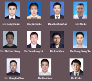
Original Article | Vol 3 | Issue 2 | July-December 2023 | page: 67-72 | RongDa Xu, JiaHui Li, ZhenCun Cai, Zhi Li, ZhiHao Liang, YuanLong Li, Lin Shen, HongLiang Tu, HongYu Zhou, Han Sun, Pei Li

DOI: https://doi.org/10.13107/jrs.2023.v03.i02.111


Author: RongDa Xu [1], JiaHui Li [1], ZhenCun Cai [1], Zhi Li [2], ZhiHao Liang [2], YuanLong Li [3], Lin Shen [4], HongLiang Tu [5], HongYu Zhou [6], Han Sun [7], Pei Li [1]

[1] Department of Orthopedics Surgery, Central Hospital Affiliated to Shenyang Medical College, Shenyang, China,
[2] Department of Orthopedics Surgery, Centro Hospitalar Conde de São Januário, Rua do Almirante Costa Cabral, Macau, China,
[3] Department of Orthopedics Surgery, Guangdong General Hospital, Yuexiu District, Guangzhou, China,
[4] Department of Hand Surgery, Central Hospital Affiliated to Shenyang Medical College, Shenyang, China,
[5] Department of Orthopedics Surgery, 242 Hospital Affiliated to Shenyang Medical College, Huanggu District, China,
[6] Department of Orthopedics Surgery, Shenyang Orthopedic Hospital, Dadong District, Shenyang, China,
[7] Department of Orthopedics Surgery, Liaoyang County Central Hospital, Liaoyang, China.

Address of Correspondence

Dr. Pei Li,
Department of Orthopedics Surgery, Central Hospital Affiliated to Shenyang Medical College, Shenyang, China.
E-mail: peili4421@gmail.com


Abstract

Objective: The objective of the study was to evaluate of the therapeutic efficacy of extracorporeal shock wave therapy (ESWT) combined with platelet-rich plasma (PRP) injection in patients with long bone fracture non-union.
Material and Methods: A total of 36 patients identified with long bone fracture non-union treated from September 2020 to September 2023 were enrolled into this study. Employing a random number table method, they were randomly divided into three groups, with 12 cases in each group. Based on the treatment modality, the groups were categorized as the ESWT group, PRP group, and combination ESWT + PRP group. Routine radiographs and musculoskeletal ultrasound were obtained before treatment and at 3-, 6-, and 9-month post-treatment intervals to observe for bone callus formation and assess fracture line imaging scores with the aim to evaluate the treatment efficacy of each group.
Results: With the extension of treatment time, the bone callus and fracture line imaging scores of the three groups gradually increased (P < 0.05). At 3-, 6-, and 9-month post-treatment, the scores of the ESWT combined with the PRP group were significantly better than those of the singular ESWT group and PRP group, and the differences were statistically significant (P < 0.05).
Conclusion: Therapy with singular ESWT, singular PRP, and combination ESWT + PRP has demonstrated effective improvement in fracture healing for patients with long bone fracture non-union. The synergistic effects of combination therapy were more significant, surpassing the efficacy of singular ESWT or PRP applications. The combined use of ESWT and PRP represents a safe and promising alternative treatment for long-bone fracture non-union, making it a compelling choice in the context of fracture healing.
Keywords: Extracorporeal shock wave therapy, Platelet-rich plasma, Non-union of fracture.


References:

1. Wu W, Tang P. The research progress on treatment methods for nonunion of bones. China Contin Med Educ 2017;9:126-9.
2. Panteli M, Vun JS, Pountos I, J Howard A, Jones E, Giannoudis PV. Biological and molecular profile of fracture non-union tissue: A systematic review and an update on current insights. J Cell Mol Med 2022;26:601-23.
3. Smolinska V, Csobonyeiova M, Zamborsky R, Danisovic L. Stem cells and their derivatives: An implication for the regeneration of nonunion fractures. Cell Transplant 2023;32:9636897231183530.
4. Wang X, Cui Y, Zhang L. Advances in treatment strategies and mechanisms for promoting fracture healing research. Chin Bull Life Sci 2021;33:121-30.
5. Schlickewei CW, Kleinertz H, Thiesen DM, Mader K, Priemel M, Frosch KH, et al. Current and future concepts for the treatment of impaired fracture healing. Int J Mol Sci 2019;20:5805.
6. Lai Y, Han J. The role and advances of extracellular vesicles in the mechanisms of bone nonunion treatment. Chin J Tissue Eng Res 2020;24:4349-55.
7. Zheng H, Zhang W, Wang Y, He B,Shen Y,Fan L. Femoral neck system combined with platelet-rich plasma in the treatment of femoral neck fracture. Chin J Tissue Eng Res 2023;27:1390-5.
8. Lv F, Li Z, Jing Y, Sun L, Li Z, Duan H. The effects and underlying mechanism of extracorporeal shockwave therapy on fracture healing. Front Endocrinol 2023;14:1188297.
9. Wang H, Shi Y. Extracorporeal shock wave treatment for post-surgical fracture nonunion: Insight into its mechanism, efficacy, safety and prognostic factors (Review). Exp Ther Med 2023;26:332.
10. Liang H, Jia H, Zhu J, Hu F, Li H, Xiao J, et al. Guidelines for extracorporeal shock wave therapy for musculoskeletal disorders in China (2023 Edition). Chin J Front Med Sci 2023;15:1-20.
11. Willems A, van der Jagt OP, Meuffels DE. Extracorporeal shock wave treatment for delayed union and nonunion fractures: A systematic review. J Orthop Trauma 2019;33:97-103.
12. Trinchese GF, Cipollaro L, Calabrese E, Maffulli N. Platelet-rich plasma, mesenchymal stem cell, and non-metallic suture-based fixation technique in a patellar fracture nonunion: A technical note and systematic review. Clin Orthop Surg 2021;13:344-51.
13. Fang J, Wang X, Jiang W, Zhu Y, Hu Y, Zhao Y, et al. Platelet-rich plasma therapy in the treatment of diseases associated with orthopedic injuries. Tissue Eng Part B Rev 2020;26:571-85.
14. Yin N, Wang Y, Ding L, Yuan J, Du L, Zhu Z, et al. Platelet-rich plasma enhances the repair capacity of muscle-derived mesenchymal stem cells to large humeral bone defect in rabbits. Sci Rep 2020;10:6771.
15. Li S, Xing F, Luo R, Liu M. Clinical effectiveness of platelet-rich plasma for long-bone delayed union and nonunion: A systematic review and meta-analysis. Front Med (Lausanne) 2022;8:771252.
16. Seabaugh KA, Thoresen M, Giguère S. Extracorporeal shockwave therapy increases growth factor release from equine platelet-rich plasma in vitro. Front Vet Sci 2017;4:205.
17. Nicholson JA, Tsang ST, MacGillivray TJ, Perks F, Simpson AH. What is the role of ultrasound in fracture management?: Diagnosis and therapeutic potential for fractures, delayed unions, and fracture-related infection. Bone Joint Res 2019;8:304-12.
18. Sun L, Zhang N, Tu H, Li P. The application value of musculoskeletal ultrasound in long tubular bone fractures and nonunion shock wave therapy. Chin J Med Ultrasound 2019;16:827-31.


How to Cite this article: Xu R, Li J, Cai Z, Li Z, Liang Z, Li Y, Shen L, Tu H, Zhou H, Sun H, Li P | Clinical Study on the Treatment of Long Bone Fracture Non-union with Extracorporeal Shock Wave Therapy Combined with Platelet-rich Plasma. | Journal of Regenerative Science | Jul-Dec 2023; 3(2): 67-72.


[Full Text HTML] [Full Text PDF] 


Very Low and Low-energetic Extracorporeal Shock Wave Treatment of Spasticity in Children and Adults – A Systematic Review

Systematic Review | Volume 2 | Issue 1 | JRS Jan – Jun 2022 | Page 03-08 | Karsten Knobloch, Henning Lohse-Busch, Andreas Gohritz, Tomas Nedelka

DOI: 10.13107/jrs.2022.v02.i01.33

Author: Karsten Knobloch [1], Henning Lohse-Busch [2], Andreas Gohritz [3], Tomas Nedelk [4,5]

[1] Department of Sports Medicine, SportPraxis Prof. Knobloch, Hannover, Germany.

[2] Zentrum für Bewegungsstörungen, Rheintalklinik Bad Krozingen, Germany.

[3] Department of Neurology, Universitätsspital Basel, Plastische, Rekonstruktive und Ästhetische Chirurgie, Handchirurgie, Suisse.

[4] Department of Neurology, Charles University in Prague, 2nd Faculty of Medicine, Prague, Czech Republic.

[5] Department of Neurology, Czech Technical University, Faculty of Biomedical Engineering, Kladno, Czech Republic.

Address of Correspondence
Prof. Dr. Karsten Knobloch,
SportPraxis Prof. Knobloch, Heiligerstr. 3, D-30159 Hannover, Germany.
E-mail: professor.knobloch@sportpraxis-knobloch.de


Abstract

Introduction: This systemic review aims to assess the modes and treatment parameters of radial and focused extracorporeal shockwave therapy (ESWT) in spasticity based on the technologies and energy levels involved.
Materials and Methods: 1086 patients from 31 randomized-controlled trials (RCT) or cohort studies are included. 300 children were studied in seven studies (3 RCTs) with two radial and four focused electromagnetic ESWT devices and 14 studies with 443 patients using focused ESWT (electrohydraulic 46 patients, electromagnetic 367 patients, and piezoelectric 30 patients).
Results: In electromagnetic focused ESWT 8 RCTs (n = 323 patients) and three cohort studies (n = 44) used either very low-energetic (0.03–0.05 mJ/mm2) with 1500–2000 shots and 4–5 Hz with 3–5 focused sessions, or low-energetic 0.07–0.12 mJ/mm2 with 1500–2000 shots with 4–5 Hz and 1–3 sessions. The 64 children in the five electromagnetic focused trials were treated with very low-energetic 0.03 mJ/mm2, 1500 shots, and three sessions. 17 studies (n = 687) were using radial technologies with 7 RCTs (n = 349) and ten cohort studies (n = 338). Among the 17 trials, four studies (1 RCT, 3 cohort studies) included 236 children treated with either very low-energetic 0.6–1 bar (two trials) or low-energetic 1.5–3 bar with 5–10 Hz. Energy-wise three radial studies were very low-energetic 0.6–1 bar and 14 studies applied low-energetic radial pressures 1.5–3 bar. Notably, the frequency was mainly 4–8 Hz in the radial studies.
Conclusion: Both, radial and focused very low- to low-energetic ESWT improve function and reduce spasticity significantly. Adverse effects were not noted with the applied very low- to low-energetic device parameters neither among children nor in adults.

Keywords: Spasticity, Extracorporeal shock wave therapy, Extracorporeal shockwave therapy, Children.


References:

1. Lohse-Busch H, Kraemer M, Reime U. A pilot investigation into the effects of extracorporeal shock waves on muscular dysfunction in children with spastic movement disorders. Schmerz 1997;11:108-12.
2. Lohse-Busch H, Kraemer M, Reime U. The use of extracorporeal shock wave fronts for treatment of muscle dysfunction of various etiologies: An overview of first results. In: Siebert, Buch , editors. Extracorporeal Shock Waves in Orthopaedics. New York, Tokyo: Springer Verlag Berlin Heidelberg; 1998. p. 215-30.
3. Riedel M, Falland R, Sailer-Kramer B, Lohse-Busch H. Komplexbehandlung mit manueller medizin und physiotherapie bei zerebral bewegungsgestörten kindern. Man Med Osteopath Med 2001;39:72-8.
4. Lohse-Busch H. Extrakorporale Stoßwellen. In: Lohse-Busch H, Riedel M, Graf Baumann T, editors. Das Therapeutische Angebot Für Bewegungsgestörte Kinder. New York: Springer Verlag Berlin Heidelberg; 2000. p. 257-74.
5. Oh JH, Park HD, Han SH, Shim GY, Choi KY. Duration of treatment effect of extracorporeal shock wave on spasticity and subgroup-analysis according to number of shocks and application site: A meta-analysis. Ann Rehabil Med 2019;43:163-77.
6. Lee JY, Kim SN, Lee IS, Jung H, Lee KS, Koh SE. Effects of extracorporeal shock wave therapy on spasticity in patients after brain injury: Ameta-analysis. J Phys Ther Sci 2014;26:1641-7.
7. Cabanas-Valdés R, Calvo-Sanz J, Urrùtia G, Serra-Llobet P, Pérez-Bellmunt A, Germán-Romero A. The effectiveness of extracorporeal shock wave therapy to reduce lower limb spasticity in stroke patients: A systematic review and meta-analysis. Top Stroke Rehabil 2020;27:137-57.
8. Xiang J, Wang W, Jiang W, Qian Q. Effects of extracorporeal shock wave therapy on spasticity in post-stroke patients: Asystematic review and meta-analysis of randomized controlled trials. J Rehabil Med 2018;50:852-9.
9. Dymarek R, Ptaszkowski K, Ptaszkowska L, Kowal M, Sopel M, Taradaj J, et al. Shock waves as a treatment modality for spasticity reduction and recovery improvement in post-stroke adults – Current evidence and qualitative systematic review. Clin Interv Aging 2020;15:9-28.
10. Liu DY, Zhong DL, Li J, Jin RJ. The effectiveness and safety of extracorporeal shock wave therapy (ESWT) on spasticity after upper motor neuron injury: Aprotocol of systematic review and meta-analysis. Medicine (Baltimore) 2020;99:e18932.
11. Corrado B, Di Luise C, Servodio Iammarrone C. Management of muscle spasticity in children with cerebral palsy by means of extracorporeal shockwave therapy: A systematic review of the literature. Dev Neurorehabil 2021;24:1-7.
12. Park DS, Kwon DR, Park GY, Lee MY. Therapeutic effect of extracorporeal shock wave therapy according to treatment session on gastrocnemius muscle spasticity in children with spastic cerebral palsy: A pilot study. Ann Rehabil Med 2015;39:914-21.
13. El-Shamy SM, Eid MA, El-Banna MF. Effect of extracorporeal shock wave therapy on gait pattern in hemiplegic cerebral palsy: A randomized controlled trial. Am J Phys Med Rehabil 2014;93:1065-72.

14. Picelli A, La Marchina E, Gajofatto F, Pontillo A, Vangelista A, Filippini R, et al. Sonographic and clinical effects of botulinum toxin Type A combined with extracorporeal shock wave therapy on spastic muscles of children with cerebral palsy. Dev Neurorehabil 2017;20:160-4.
15. Amelio E, Manganotti P. Effect of shock wave stimulation on hypertonic plantar flexor muscles in patients with cerebral palsy: A placebo-controlled study. J Rehabil Med 2010;42:339-43.
16. Lin Y, Wang G, Wang B. Rehabilitation treatment of spastic cerebral palsy with radial extracorporeal shock wave therapy and rehabilitation therapy. Medicine (Baltimore) 2018;97:e13828.
17. Wang T, Du L, Shan L, Dong H, Feng J, Kiessling MC, et al. A prospective case-control study of radial extracorporeal shock wave therapy for spastic plantar flexor muscles in very young children with cerebral palsy. Medicine (Baltimore) 2016;95:e3649.
18. Mirea A, Onose G, Padure L, Rosulescu E. Extracorporeal shockwave therapy (ESWT) benefits in spastic children with cerebral palsy (CP). J Med Life 2014;7:127-32.
19. Gonkova MI, Ilieva EM, Ferriero G, Chavdarov I. Effect of radial shock wave therapy on muscle spasticity in children with cerebral palsy. Int J Rehabil Res 2013;36:284-90.
20. Santamato A, Micello MF, Panza F, Fortunato F, Logroscino G, Picelli A, et al. Extracorporeal shock wave therapy for the treatment of poststroke plantar-flexor muscles spasticity: A prospective open-label study. Top Stroke Rehabil 2014;21 Suppl 1:S17-24.
21. Sohn MK, Cho KH, Kim YJ, Hwang SL. Spasticity and electrophysiologic changes after extracorporeal shock wave therapy on gastrocnemius. Ann Rehabil Med 2011;35:599-604.
22. Lee CH, Lee SH, Yoo JI, Lee SU. Ultrasonographic evaluation for the effect of extracorporeal shock wave therapy on gastrocnemius muscle spasticity in patients with chronic stroke. PM R 2019;11:363-71.
23. Wu YT, Yu HK, Chen LR, Chang CN, Chen YM, Hu GC. Extracorporeal shock waves versus botulinum toxin type A in the treatment of poststroke upper limb spasticity: A randomized noninferiority trial. Arch Phys Med Rehabil 2018;99:2143-50.
24. Taheri P, Vahdatpour B, Mellat M, Ashtari F, Akbari M. Effect of extracorporeal shock wave therapy on lower limb spasticity in stroke patients. Arch Iran Med 2017;20:338-43.
25. Yoon SH, Shin MK, Choi EJ, Kang HJ. Effective site for the application of extracorporeal shock-wave therapy on spasticity in chronic stroke: Muscle belly or myotendinous junction. Ann Rehabil Med 2017;41:547-55.
26. Santamato A, Notarnicola A, Panza F, Ranieri M, Micello MF, Manganotti P, et al. SBOTE study: Extracorporeal shock wave therapyversus electrical stimulation after botulinum toxin type a injection for post-stroke spasticity – A prospective randomized trial. Ultrasound Med Biol 2013;39:283-91.
27. Manganotti P, Amelio E. Long-term effect of shock wave therapy on upper limb hypertonia in patients affected by stroke. Stroke 2005;36:1967-71.
28. Troncati F, Paci M, Myftari T, Lombardi B. Extracorporeal Shock Wave Therapy reduces upper limb spasticity and improves motricity in patients with chronic hemiplegia: A case series. NeuroRehabilitation 2013;33:399-405.
29. Moon SW, Kim JH, Jung MJ, Son S, Lee JH, Shin H, et al. The effect of extracorporeal shock wave therapy on lower limb spasticity in subacute stroke patients. Ann Rehabil Med 2013;37:461-70.
30. Radinmehr H, Ansari NN, Naghdi S, Tabatabaei A, Moghimi E. Comparison of therapeutic ultrasound and radial shock wave therapy in the treatment of plantar flexor spasticity after stroke: A prospective, single-blind, randomized clinical trial. J Stroke Cerebrovasc Dis 2019;28:1546-54.
31. Marinelli L, Mori L, Solaro C, Uccelli A, Pelosin E, Currà A, et al. Effect of radial shock wave therapy on pain and muscle hypertonia: A double-blind study in patients with multiple sclerosis. Mult Scler 2015;21:622-9.
32. Vidal X, Morral A, Costa L, Tur M. Radial extracorporeal shock wave therapy (rESWT) in the treatment of spasticity in cerebral palsy: A randomized, placebo-controlled clinical trial. NeuroRehabilitation 2011;29:413-9.
33. Li TY, Chang CY, Chou YC, Chen LC, Chu HY, Chiang SL, et al. Effect of radial shock wave therapy on spasticity of the upper limb in patients with chronic stroke: A prospective, randomized, single blind, controlled trial. Medicine (Baltimore) 2016;95:e3544.
34. Wu YT, Chang CN, Chen YM, Hu GC. Comparison of the effect of focused and radial extracorporeal shock waves on spastic equinus in patients with stroke: A randomized controlled trial. Eur J Phys Rehabil Med 2018;54:518-25.
35. Dymarek R, Taradaj J, Rosińczuk J. The effect of radial extracorporeal shock wave stimulation on upper limb spasticity in chronic stroke patients: A single-blind, randomized, placebo-controlled study. Ultrasound Med Biol 2016;42:1862-75.
36. Megna M, Marvulli R, Farì G, Gallo G, Dicuonzo F, Fiore P, et al. Pain and muscles properties modifications after botulinum toxin type A (BTX-A) and radial extracorporeal shock wave (rESWT) combined treatment. Endocr Metab Immune Disord Drug Targets 2019;19:1127-33.
37. Kim YW, Shin JC, Yoon JG, Kim YK, Lee SC. Usefulness of radial extracorporeal shock wave therapy for the spasticity of the subscapularis in patients with stroke: Apilot study. Chin Med J (Engl) 2013;126:4638-43.
38. Sawan S, Abd-Allah F, Hegazy MM, Farrag MA, El-Den NH. Effect of shock wave therapy on ankle plantar flexors spasticity in stroke patients. NeuroRehabilitation 2017;40:115-8.
39. Radinmehr H, Nakhostin Ansari N, Naghdi S, Olyaei G, Tabatabaei A. Effects of one session radial extracorporeal shockwave therapy on post-stroke plantarflexor spasticity: A single-blind clinical trial. Disabil Rehabil 2017;39:483-90.
40. Kim TG, Bae SH, Kim GY, Kim KY. The effects of extracorporeal shock wave therapy on stroke patients with plantar fasciitis. J Phys Ther Sci 2015;27:523-6.
41. Dymarek R, Taradaj J, Rosińczuk J. Extracorporeal shock wave stimulation as alternative treatment modality for wrist and fingers spasticity in poststroke patients: A prospective, open-label, preliminary clinical trial. Evid Based Complement Alternat Med 2016;2016:4648101.
42. Daliri SS, Forogh B, Emami Razavi SZ, Ahadi T, Madjlesi F, Ansari NN. A single blind, clinical trial to investigate the effects of a single session extracorporeal shock wave therapy on wrist flexor spasticity after stroke. NeuroRehabilitation 2015;36:67-72.
43. Amelio E, Manganotti P. Effect of Shock Wave Therapy in Patients Affected by Stroke with Upper Limb Spasticity: Neurophysiologic and Clinical Study. International Federation of Societies for Surgery of the Hand; 2004. p. 535-40.
44. Chaussy C, Brendel W, Schmiedt E. Extracorporeally induced destruction of kidney stones by shock waves. Lancet 1980;2:1265-8.
45. Louwerens JK, Sierevelt IN, Kramer ET, Boonstra R, van den Bekerom MP, van Royen BJ, et al. Comparing ultrasound-guided needling combined with a subacromial corticosteroid injection versus high-energy extracorporeal shockwave therapy for calcific tendinitis of the rotator cuff: A randomized controlled trial. Arthroscopy 2020;36:1823-33.e1.
46. Wang YC, Chen SJ, Huang PJ, Huang HT, Cheng YM, Shih CL. Efficacy of different energy levels used in focused and radial extracorporeal shockwave therapy in the treatment of plantar fasciitis: A meta-analysis of randomized placebo-controlled trials. J Clin Med 2019;8:1497.


How to Cite this article: Knobloch K, Lohse-Busch H, Gohritz A, Nedelka T | Very Low and Low-energetic Extracorporeal Shock Wave Treatment of Spasticity in Children and Adults–A Systematic Review. | Journal of Regenerative Science | Jan – Jun 2022; 2(1): 03-08.

[Full Text HTML] [Full Text PDF] [XML]