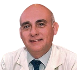
China, The Awakened Giant

Editorial | Vol 3 | Issue 2 |  July-December 2023 | page: 01-02 | Daniel Moya

DOI: https://doi.org/10.13107/jrs.2023.v03.i02.85


Author: Daniel Moya [1]

[1] Department of Orthopaedics. Buenos Aires British Hospital, Argentina.


Address of Correspondence
Dr. Daniel Moya,
Department of Orthopaedics. Buenos Aires British Hospital, Argentina.
E-mail: drdanielmoya@yahoo.com.ar


Editorial:

The history of Chinese medicine is as long and legendary as that of the country itself. Its origins date back 3000 years [1]. Scientific knowledge and medical practice has gone through numerous stages.
Between the 8th and 3rd centuries BC, China went through a period of great cultural and intellectual development called the “Hundred Schools of Thought” [2]. An attempt was made to seek the explanation of the phenomena of the universe in nature itself, leaving aside explanations based on magic and myths [2]. The new ideas discussed and developed during this period have profoundly influenced philosophical views and lifestyles up to the present day in East Asian countries.
A fundamental milestone was the publication of “Huangdi Neijing“. Also known as the “Inner Canon of Huangdi“ or “Inner Canon of the Yellow Emperor“, it is the earliest surviving work on Chinese medicine[3]. Its author, Emperor Huangdi, is not only considered the initiator of Chinese Traditional Medicine but also the father of Chinese civilization.
Caring for the health of the population has been a priority in this country throughout the centuries. As a history lover, it is difficult for me to find another example of a nation embarking on an unequal war to defend its public health. This happened when the Western powers, led by the British Empire, sought to create opium addiction among the Chinese population in order to balance their trade balance by trafficking drugs [4]. These infamous conflicts went down in history as the “Opium Wars” and cost the lives of thousands of Chinese citizens.
In the last decades, China’s healthcare system has made great achievements in the management of medical services and public health for the Chinese people[5]. Average life expectancy at birth was 35 years before the founding of new China, and it reached 77.0 years in 2018 [6]. The projected life expectancy at birth in mainland China in 2035 is 81,3 years [7].
On April 6, 2009, China presented an action plan to undertake a radical and ambitious reform of the health system [8]. The goal is to achieve universal health coverage.
The history of shock waves in this country is a reflection of the described dynamism and historical background. The beginnings of its use in China can be traced back to the 1980s in the urological field. Prof. Xing Gengyan was the pioneer of indications in musculoskeletal pathology starting in 1993. Since that moment he has been a promoter not only of research and clinical applications, but also of medical education. In 2019, he organized in Beijing the largest international shock wave congress in history. It is a great honor that he has contributed to this issue by writing about the development of this therapeutic practice in China.
In parallel with healthcare and academic development, the Chinese industry has risen to the occasion by providing high-quality devices.
The content of this volume reflects not only the ability of our Chinese colleagues but also their openness to the world and their generosity in sharing information. Numerous universities and hospitals from different regions throughout China have collaborated selflessly. This could not have been accomplished without the monumental task of Dr. Sun Wei, our Guest Editor.
Napoleon Bonaparte is credited with the phrase “China is a sleeping giant, when she wakes she will shake the world“. Two hundred years later the giant is awake and brimming with energy.


References

[1] Reyes G Ariel E. Evolución Histórica de la Medicina Tradicional China. Comunidad y Salud [Internet]. 2008 Dic [citado 2023 Dic 27] ; 6( 2 ): 42-49. Disponible en: http://ve.scielo.org/scielo.php?script=sci_arttext&pid=S1690-32932008000200005&lng=es
[2] Orígenes de la Medicina China. Escuela Li Ping de acupuntura y Medicina Tradicional China. chrome-extension://efaidnbmnnnibpcajpcglclefindmkaj/https://escuelaliping.com/wp-content/uploads/2013/10/Tema1.pdf
[3] Huang di nei jing su wen. Library of the congress. https://www.loc.gov/item/2021666312
[4] Travis Hanes III W, and Sanello F. The Addiction of One Empire and the Corruption of Another. Ed. Sourcebooks. 2004. ISBN-13 ‏ : ‎ 978-1402201493
[5] Chen C, Liu M. Achievements and Challenges of the Healthcare System in China. Cureus. 2023 May 15;15(5):e39030. doi: 10.7759/cureus.39030. PMID: 37378106; PMCID: PMC10292030.
[6] Yuan, X., Gao, Y. Demographic transition and economic miracles in China: an analysis based on demographic perspective. IJEPS 14, 25–45 (2020). https://doi.org/10.1007/s42495-019-00030-0
[7] Bai R, Liu Y, Zhang L, Dong W, Bai Z, Zhou M. Projections of future life expectancy in China up to 2035: a modelling study. Lancet Public Health. 2023 Dec;8(12):e915-e922. doi: 10.1016/S2468-2667(22)00338-3. Epub 2023 Mar 30. PMID: 37004714; PMCID: PMC10188127.
[8]China’s latest revolution: Basic health care for all. https://www.ilo.org/global/about-the-ilo/mission-and-objectives/features/WCMS_188582/lang–en/index.htm


How to Cite this article: Moya D | China, The Awakened Giant. | Journal of Regenerative Science | Jul-Dec 2023; 3(2): 01-02.


  (Abstract    Full Text HTML)   (Download PDF)


The History of Shock Wave Medicine Development in China

Editorial | Vol 3 | Issue 2 |  July-December 2023 | page: 03-04 | Shuitao Liu, Gengyan Xing

DOI: https://doi.org/10.13107/jrs.2023.v03.i02.87


Author: Shuitao Liu [1], Gengyan Xing [2]

[1] Department of Traumatic Orthopedics, Armed Police Characteristic Medical Center., Tianjin, China,
[2] Corresponding author: Gengyan Xing, Department of Orthopedic, The Third Medical Center of Chinese People’s Liberation Army General Hospital, Beijing, China.

Address of Correspondence
Dr. Gengyan Xing,
Department of Orthopedic, The Third Medical Center of Chinese People’s Liberation Army General Hospital, Beijing,
China.
E-mail: xgy1350138@163.com


Editorial

The development of shock wave medicine in China can be traced back to the 1980s. At that time, shock waves were applied to treat urinary tract stones with good results. In 1993, Professor Xing Gengyan pioneered the application of extracorporeal shock wave therapy for humeral epicondylitis, marking a significant milestone in the advancement of shock wave medicine in China. With the deepening of research on shock waves and technological advancements, the indications for shock wave medicine have been continuously expanded, achieving good results in treating bone tissue diseases such as delayed fracture healing and non-union. To further promote the experience of shock wave therapy, Professor Xing Gengyan successively held 8 national continuing education programs on “Extracorporeal Shock Wave Therapy for Bone and Muscle Disorders,” training thousands of doctors who mastered shock wave therapy techniques. With the conduct of numerous clinical studies and accumulation of data, the China National Medical Products Administration approved the domestic production of extracorporeal shock wave therapy machines for treating orthopedic diseases in August 2000, marking the official entry of shock wave medicine into a high-speed development phase in China.
As clinical applications continue to advance and expand, Chinese scholars have begun to explore the mechanisms of action of shock waves. In 2004, Professor Xing Ganyan’s research on “Osteoblast Mechanochemical Signal Transduction and Related Gene Expression Following ESWT” was funded by the National Natural Science Foundation of China, resulting in a wealth of published findings. In 2007, Professor Xing Ganyan edited and published the first monograph on shock wave medicine, <Extracorporeal Shock Wave Therapy for Bone and Muscle Diseases (First Edition)>, which consists of two parts and eight chapters. This comprehensive summary of over a decade of clinical application experience and foundational research laid the foundation for shock wave medicine in China.
With the rapid advancement of China’s medical level, Chinese scholars’ research on shock waves has gradually gained global recognition. A significant number of papers on shock waves published by Chinese scholars has been indexed, and their research achievements have been communicated at international conferences. Concurrently, as the depth of research on shock wave therapy increases and instruments advance, shock waves are no longer confined to treating urinary tract stones, non-unions, osteonecrosis, and tendinopathy [1-4]. They have also achieved promising results in treating myocardial infarction, skin ulcers, tumors, nerve injuries, and male dysfunction, among others.
In December 2013, the Chinese Shock Wave Medicine Professional Committee was established in Beijing, with Professor Xing Gengyan serving as the inaugural Chairman. Since then, shock wave medicine has had an independent academic organization. Building on past experiences, in 2014, the <Expert Consensus on Extracorporeal Shock Wave Therapy for Bone and Muscle Diseases> was released [5], significantly advancing the scientific and standardized development of shock wave medicine. In 2015, the <Extracorporeal Shock Wave Therapy for Bone and Muscle Diseases (Second Edition)> was published, expanding the volume to 5 articles and 19 chapters, with a significant addition of numerous recent research findings. In July 2016, at the annual meeting of the international society for medical shock wave treatment (ISMST) held in Malaysia, Professor Xing Gengyan was elected as the President of the ISMST. China secured the hosting rights for the 22nd annual conference in 2019, signifying that China is at the forefront of shock wave medicine development worldwide.
The year 2019 marked the most memorable year in the history of shock wave development in Chinese medicine. Building upon the previous two editions of the <Expert Consensus on Extracorporeal Shock Wave Therapy for Bone and Muscle Diseases>, Chinese scholars integrated evidence-based medical experiences and released the <Chinese Guidelines for Extracorporeal Shock Wave Therapy for Bone and Muscle Diseases (2019 Edition) [6], thereby standardizing and scientifically advancing the application of shock waves in China. In May of the same year, the ISMST 22nd International Congress on Medical Shock Waves convened in Beijing, attended by over 1,200 experts and scholars from nearly 30 countries. This was the largest-attended conference in the history of shock wave medicine, covering the broadest range of academic interests and delving into the most in-depth discussions on various research topics, significantly advancing the development of shock wave medicine in China.
Currently, over 3,000 hospitals in China possess extracorporeal shock wave therapy systems, covering treatment fields such as orthopedics, urology, plastic surgery, cardiology, stomatology, oncology, and rehabilitation, treating millions of patients annually [7-9]. Concurrently, Chinese scholars in the field of shock wave therapy are continually innovating, integrating extracorporeal shock waves with techniques such as arthroscopy, stem cells, and nanomaterials to achieve synergistic therapeutic effects, resulting in favorable outcomes [10-12]. Each year, over 500 related research papers are published.
Further, elucidating the biological mechanisms of shock waves in clinical efficacy at the organizational, cellular, and molecular levels will continuously expand the application scope of shock waves. In response to the relative lack of high-level evidence for shock wave research, the China Shock Wave Medical Association is organizing multi-center, large-sample clinical studies; standardized training for practitioners and accreditation of treatment institutions have also been put on the agenda. It is believed that in the near future, China’s shock wave medicine will witness a richer array of research achievements, bringing blessings to more patients.


References:

1. Xing G. The past, present and future of shockwave medicine. Chin J Front Med Sci 2014;6:1-2.
2. Wang Y, Guo T, Cai HY, Ma TK, Tao SM, Sun S, et al. Cardiac shock wave therapy reduces angina and improves myocardial function in patients with refractory coronary artery disease. Clin Cardiol 2010;33:693-9.
3. Liu J, Zhou F, Li GY, Wang L, Li HX, Bai GY, et al. Evaluation of the effect of different doses of low energy shock wave therapy on the erectile function of streptozotocin (STZ)-induced diabetic rats. Int J Mol Sci 2013;14:10661-73.
4. Yan X, Yang G, Cheng L, Chen M, Cheng X, Chai Y, et al. Effect of extracorporeal shock wave therapy on diabetic chronic wound healing and its histological features. Zhongguo Xiu Fu Chong Jian Wai Ke Za Zhi 2012;26:961-7.
5. Xing G, Huang C, Huang Z, Li Y, Li Z, Liu Y, et al. Professional Committee of Shock Wave Medicine of Chinese Research Hospital Association. Expert consensus of extracorporeal shock wave therapy for bone and muscle diseases. Chin J Front Med Sci 2014;6:170-7.
6. Xing G, Zhang H, Liu S, Zhao Z. Chinese guidelines for extracorporeal shockwave therapy for bone and muscle diseases (2019 Edition). Chin J Front Med Sci 2019;11:1-10.
7. Xu Y, Wu K, Liu Y, Geng H, Zhang H, Liu S, et al. The effect of extracorporeal shock wave therapy on the treatment of moderate to severe knee osteoarthritis and cartilage lesion. Medicine (Baltimore) 2019;98:e15523.
8. Li F, Zhen Z, Sun SJ, Jiang Y, Liang WH, Belau M, et al. Attenuation of myocardial dysfunction in hypertensive cardiomyopathy using non-R-wave-synchronized cardiac shock wave therapy. Int J Mol Sci 2022;23:13274.
9. Huang Q, Yan P, Xiong H, Shuai T, Liu J, Zhu L, et al. Extracorporeal shock wave therapy for treating foot ulcers in adults with type 1 and type 2 diabetes: A systematic review and meta-analysis of randomized controlled trials. Can J Diabetes 2020;44:196-204.e3.
10. Liang H, Chen K, Xie J, Yao L, Liu Y, Hu F, et al. A bone-penetrating precise controllable drug release system enables localized treatment of osteoporotic fracture prevention via modulating osteoblast-osteoclast communication. Small 2023;19:e2207195.
11. Bai X, Gao Y, Zhang M, Chang YN, Chen K, Li J, et al. Carboxylated gold nanoparticles inhibit bone erosion by disturbing the acidification of an osteoclast absorption microenvironment. Nanoscale 2020;12:3871-8.
12. Zeng L, Geng H, Gu W, Ma S, Qin Y, Xia S, et al. Au nanoparticles attenuate RANKL-induced osteoclastogenesis by suppressing pre-osteoclast fusion. J Nanosci Nanotechnol 2019;19:2166-73.


How to Cite this article: Liu S, Xing G | The History of Shock Wave Medicine Development in China. | Journal of Regenerative Science | Jul-Dec 2023; 3(2): 03-04.

[Full Text HTML] [Full Text PDF] 


Shock Wave Medicine: A Transformative Evolution in Modern Medicine

Original Article | Vol 3 | Issue 2 |  July-December 2023 | page: 05-09 | Sunte Li, Xiaoyu Fan, Wei Sun

DOI: https://doi.org/10.13107/jrs.2023.v03.i02.89


Author: Sunte Li [1], Xiaoyu Fan [2], Wei Sun [3, 4]

[1] Friends Central School, Philadelphia, Pennsylvania, USA,
[2] Department of Surgery, Peking University People’s Hospital, Beijing, China,
[3] Department of Orthopaedic Surgery, Perelman School of Medicine, University of Pennsylvania, Philadelphia, Pennsylvania, USA,
[4] Department of Orthopedics, Shockwave Center, China-Japan Friendship Hospital, Chaoyang, Beijing, China.

Address of Correspondence
Dr. Wei Sun,
Department of Orthopaedic Surgery, Perelman School of Medicine, University of Pennsylvania, Philadelphia, Pennsylvania, USA/Department of Orthopedics, Shockwave Center, China-Japan Friendship Hospital, Chaoyang, Beijing, China.
E-mail: wei.sun@pennmedicine.upenn.edu


Abstract

Since its inception as extracorporeal shock wave lithotripsy in the 1980s, the landscape of medical treatment has been revolutionized by the evolution of shock wave therapy. Over four decades, this therapy, now known as extracorporeal shock wave therapy (ESWT), has emerged as a cornerstone in modern medicine, redefining treatment paradigms across various medical disciplines. Certainly, despite the promising outcomes witnessed in various medical conditions such as musculoskeletal disorders, wound healing, urinary calculi, and erectile dysfunction,
it is crucial to acknowledge that shock wave therapy’s relatively short clinical tenure necessitates a cautious approach. While its effectiveness has been repeatedly demonstrated, establishing industry-standard protocols through large-scale, prospective randomized controlled trials remains imperative to solidify its standing in medical practice.
The integration of Artificial Intelligence technology holds significant promise for the future of shockwave medicine, enabling personalized treatment plans, real-time feedback, and improved cost-effectiveness.
Keywords: Shock waves, ESWT, Shockwave


References:

1. Seoane LM, Salvador JB, Alba A, Fentes DA. Technological innovations in shock wave lithotripsy. Actas Urol Esp (Engl Ed) 2024; (48)-1:105-110. https://doi.org/10.1016/j.acuroe.2023.09.001
2. Moya D, Ramón S, Schaden W, Wang CJ, Guiloff L, Cheng JH. The role of extracorporeal shockwave treatment in musculoskeletal disorders. J Bone Joint Surg Am 2018;100:251-63.
3. Porst H. Review of the current status of low intensity extracorporeal shockwave therapy (Li-ESWT) in erectile dysfunction (ED), Peyronie’s disease (PD), and sexual rehabilitation after radical prostatectomy with special focus on technical aspects of the different marketed ESWT devices including personal experiences in 350 patients. Sex Med Rev 2021;9:93-122.
4. Van der Worp H, Van den Akker-Scheek I, Van Schie H, Zwerver J. ESWT for tendinopathy: Technology and clinical implications. Knee Surg Sports Traumatol Arthrosc 2013;21:1451-8.
5. Schroeder AN, Tenforde AS, Jelsing EJ. Extracorporeal shockwave therapy in the management of sports medicine injuries. Curr Sports Med Rep 2021;20:298-305.
6. Wang H, Shi Y. Extracorporeal shock wave treatment for post-surgical fracture nonunion: Insight into its mechanism, efficacy, safety and prognostic factors (Review). Exp Ther Med 2023;26:332.
7. Simplicio CL, Purita J, Murrell W, Santos GS, Dos Santos RG, Lana JF. Extracorporeal shock wave therapy mechanisms in musculoskeletal regenerative medicine. J Clin Orthop Trauma 2020;11:S309-18.
8. Wigley CH, Janssen TJ, Mosahebi A. Shock wave therapy in plastic surgery: A review of the current indications. Aesthet Surg J 2023;43:370-86.
9. Kuo YR, Wang CT, Wang FS, Chiang YC, Wang CJ. Extracorporeal shock-wave therapy enhanced wound healing via increasing topical blood perfusion and tissue regeneration in a rat model of STZ-induced diabetes. Wound Repair Regen 2009;17:522-30.
10. Lee SY, Joo SY, Cho YS, Hur GY, Seo CH. Effect of extracorporeal shock wave therapy for burn scar regeneration: A prospective, randomized, double-blinded study. Burns 2021;47:821-7.
11. Yao H, Wang X, Liu H, Sun F, Tang G, Bao X et al. Systematic Review and Meta-Analysis of 16 Randomized Controlled Trials of Clinical Outcomes of Low-Intensity Extracorporeal Shock Wave Therapy in Treating Erectile Dysfunction. Am J Mens Health. 2022 Mar-Apr;16(2):15579883221087532. doi: 10.1177/15579883221087532. PMID: 35319291; PMCID: PMC8949743.
12. Dong L, Chang D, Zhang X, Li J, Yang F, Tan K, et al. Effect of low-intensity extracorporeal shock wave on the treatment of erectile dysfunction: A systematic review and meta-analysis. Am J Mens Health 2019;13:2. Published online. Open access: https://journals.sagepub.com/action/showCitFormats?doi=10.1177%2F1557988319846749&mobileUi=0
13. Wu WL, Bamodu OA, Wang YH, Hu SW, Tzou KY, Yeh CT, et al. Extracorporeal shockwave therapy (ESWT) alleviates pain, enhances erectile function and improves quality of Life in patients with chronic prostatitis/chronic pelvic pain syndrome. J Clin Med 2021;3602.
14. Radu CA, Kiefer J, Horn D, Rebel M, Koellensperger E, Gebhard MM, et al. Shock wave treatment in composite tissue allotransplantation. Eplasty 2011;11:e37.
15. Li HX, Zhang ZC, Peng J. Low-intensity extracorporeal shock wave therapy promotes recovery of sciatic nerve injury and the role of mechanical sensitive YAP/TAZ signaling pathway for nerve regeneration. Chin Med J (Engl) 2021;134:2710-20.
16. Mittermayr R, Hartinger J, Antonic V, Meinl A, Pfeifer S, Stojadinovic A, et al. Extracorporeal shock wave therapy (ESWT) minimizes ischemic tissue necrosis irrespective of application time and promotes tissue revascularization by stimulating angiogenesis. Ann Surg 2011;253:1024-32.
17. Yamaya S, Ozawa H, Kanno H, Kishimoto KN, Sekiguchi A, Tateda S, et al. Low-energy extracorporeal shock wave therapy promotes vascular endothelial growth factor expression and improves locomotor recovery after spinal cord injury. J Neurosurg 2014;121:1514-25.
18. López-Marín LM, Rivera AL, Fernández F, Loske AM. Shock wave-induced permeabilization of mammalian cells. Phys Life Rev 2018;26-27:1-38.
19. Yeh KH, Sheu JJ, Lin YC, Sun CK, Chang LT, Kao YH, et al. Benefit of combined extracorporeal shock wave and bone marrow-derived endothelial progenitor cells in protection against critical limb ischemia in rats. Crit Care Med 2012;40:169-77.
20. Reichenberger MA, Heimer S, Schaefer A, Lass U, Gebhard MM, Germann G, et al. Extracorporeal shock wave treatment protects skin flaps against ischemia-reperfusion injury. Injury 2012;43:374-80.
21. Sung PH, Fu M, Chiang HJ, Huang CR, Chu CH, Lee MS, et al. Reduced effects of cardiac extracorporeal shock wave therapy on angiogenesis and myocardial function recovery in patients with end-stage coronary artery and renal diseases. Biomed J 2021;44:S201-9.
22. Oktaş B, Orhan Z, Erbil B, Değirmenci E, Ustündağ N. Effect of extracorporeal shock wave therapy on fracture healing in rat femural fractures with intact and excised periosteum. Eklem Hastalik Cerrahisi 2014;25:158-62.
23. Qiao HY, Xin L, Wu SL. Analgesic effect of extracorporeal shock-wave therapy for frozen shoulder: A randomized controlled trial protocol. Medicine (Baltimore) 2020;99:e21399.
24. Fiani B, Davati C, Griepp DW, Lee J, Pennington E, Moawad CM. Enhanced spinal therapy: Extracorporeal shock wave therapy for the spine. Cureus 2020;12:e11200.
25. Özkan E, Şenel E, Bereket MC, Önger ME. The effect of shock waves on mineralization and regeneration of distraction zone in osteoporotic rabbits. Ann Med 2023;55:1346-54.
26. Shi L, Gao F, Sun W, Wang B, Guo W, Cheng L, et al. Short-term effects of extracorporeal shock wave therapy on bone mineral density in postmenopausal osteoporotic patients. Osteoporos Int 2017;28:2945-53.
27. Hao L, Liu Y, Wang T, Guo HL, Wang D, Bi YW, et al. Extracorporeal shock wave lithotripsy is safe and effective for geriatric patients with chronic pancreatitis. J Gastroenterol Hepatol 2019;34:466-73.
28. Klang E, Portugez S, Gross R, Lerner KR, Brenner A, Gilboa M, et al. Advantages and pitfalls in utilizing artificial intelligence for crafting medical examinations: A medical education pilot study with GPT-4. BMC Med Educ 2023;23:772.
29. Wójcik S, Rulkiewicz A, Pruszczyk P, Lisik W, Poboży M, Domienik-Karłowicz J. Beyond ChatGPT: What does GPT-4 add to healthcare? The dawn of a new era. Cardiol J 2023;30:1018-25.
30. Mun C, Ha H, Lee O, Cheon M. Enhancing AI-CDSS with U-AnoGAN: Tackling data imbalance. Comput Methods Programs Biomed 2023;244:107954.
31. Palavicini G. Intelligent health: Progress and benefit of artificial intelligence in sensing-based monitoring and disease diagnosis. Sensors (Basel) 2023;23:9053.


How to Cite this article: Li S, Fan X, Sun W. | Shock Wave Medicine: A Transformative Evolution in Modern Medicine. | Journal of Regenerative Science | Jul-Dec 2023; 3(2): 05-09.


[Full Text HTML] [Full Text PDF] 


Extracorporeal Shockwave Therapy in Osteonecrosis of the Femoral Head: Where Do We Stand?

Original Article | Vol 3 | Issue 2 |  July-December 2023 | page: 10-13 | Tianyang Liu, Fuqiang Gao, Wei Sun

DOI: https://doi.org/10.13107/jrs.2023.v03.i02.91


Author: Tianyang Liu [1], Fuqiang Gao [2], Wei Sun [2, 3]

[1] Capital Medical University China-Japanese Friendship Clinical Medical Research Institute, Beijing, China,
[2] Department of Orthopedics, Shockwave Center, China-Japan Friendship Hospital, Chaoyang, Beijing, China,
[3] Department of Orthopaedic Surgery, Perelman School of Medicine, University of Pennsylvania, Philadelphia, Pennsylvania, USA.

Address of Correspondence
Dr. Wei Sun,
Department of Orthopedics, Shockwave Center, China-Japan Friendship Hospital, Chaoyang, Beijing,
China/Department of Orthopaedic Surgery, Perelman School of Medicine, University of Pennsylvania, Philadelphia, Pennsylvania, USA.
E-mail: wei.sun@pennmedicine.upenn.edu


Abstract

Osteonecrosis of the femoral head (ONFH) is a progressive disease characterized by ischemic lesions and structural damage in the head of the femur caused by insufficient blood supply due to multiple etiologies. As a safe, effective, non-invasive, and low-cost treatment strategy, Extracorporeal Shockwave Treatment (ESWT) is now widely applied in musculoskeletal disorders such as delayed bone healing, bone marrow edema (BME), knee osteoarthritis, and certain types of avascular bone necrosis. ESWT may promote vascularization and osteogenesis through a cascade reaction stimulated by the transformation of physical energy, promoting tissue regeneration, and repair. ESWT is recommended in treating early-stage ONFH.
Keywords: Shock waves; ESWT, Femoral head osteonecrosis


References:

1. Mont MA, Salem HS, Piuzzi NS, Goodman SB, Jones LC. Nontraumatic osteonecrosis of the femoral head: Where do we stand today? A 5-year update. J Bone Joint Surg Am 2020;102:1084-99.
2. Zhang Q, Liu L, Sun W, Gao F, Cheng L, Li Z. Extracorporeal shockwave therapy in osteonecrosis of femoral head: A systematic review of now available clinical evidences. Medicine (Baltimore) 2017;96:e5897.
3. Haupt G. Use of extracorporeal shock waves in the treatment of pseudarthrosis, tendinopathy and other orthopedic diseases. J Urol 1997;158:4-11.
4. Auersperg V, Trieb K. Extracorporeal shock wave therapy: An update. EFORT Open Rev 2020;5:584-92.
5. Moya D, Ramón S, Schaden W, Wang CJ, Guiloff L, Cheng JH. The role of extracorporeal shockwave treatment in musculoskeletal disorders. J Bone Joint Surg Am 2018;100:251-63.
6. Wang QW, Zhang QY, Gao FQ, Sun W. Focused extra-corporeal shockwave treatment during early stage of osteonecrosis of femoral head. Chin Med J (Engl) 2019;132:1867-9.
7. Cheng JH, Wang CJ. Biological mechanism of shockwave in bone. Int J Surg 2015;24:143-6.
8. Wu X, Wang Y, Fan X, Xu X, Sun W. Extracorporeal shockwave relieves endothelial injury and dysfunction in steroid-induced osteonecrosis of the femoral head via miR-135b targeting FOXO1: In vitro and in vivo studies. Aging (Albany NY) 2022;14:410-29.
9. Li B, Wang R, Huang X, Ou Y, Jia Z, Lin S, et al. Extracorporeal shock wave therapy promotes osteogenic differentiation in a rabbit osteoporosis model. Front Endocrinol (Lausanne) 2021;12:627718.
10. Hsu SL, Jhan SW, Hsu CC, Wu YN, Wu KL, Kuo CA, et al. Effect of three clinical therapies on cytokines modulation in the hip articular cartilage and bone improvement in rat early osteonecrosis of the femoral head. Biomed J 2022;46:100571.
11. Sun W, Li Z. Extracorporeal shockwave therapy for osteonecrosis of femoral head: Traps and challenges. Zhongguo Xiu Fu Chong Jian Wai Ke Za Zhi 2019;33:659-61.
12. Ludwig J, Lauber S, Lauber HJ, Dreisilker U, Raedel R, Hotzinger H. High-energy shock wave treatment of femoral head necrosis in adults. Clin Orthop Relat Res 2001:119-26.
13. Qu RD, Sun W. Interpretation of international society for medical shockwave treatment consensus-2021 in diagnosis and treatment of musculoskeletal diseases. Chin J Gen Pract 2022;21:826-30.
14. Gao F, Sun W, Li Z, Guo W, Wang W, Cheng L, et al. High-energy extracorporeal shock wave for early stage osteonecrosis of the femoral head: A single-center case series. Evid Based Complement Alternat Med 2015;2015:468090.
15. Sun W, Gao F, Guo W, Wang B, Li Z, Cheng L, et al. Focused extracorporeal shock wave for osteonecrosis of the femoral head with leukemia after allo-HSCT: A case series. Bone Marrow Transplant 2016;51:1507-9.
16. Yang X, Shi L, Zhang T, Gao F, Sun W, Wang P, et al. High-energy focused extracorporeal shock wave prevents the occurrence of glucocorticoid-induced osteonecrosis of the femoral head: A prospective randomized controlled trial. J Orthop Translat 2022;36:145-51.
17. Xie K, Mao Y, Qu X, Dai K, Jia Q, Zhu Z, et al. High-energy extracorporeal shock wave therapy for nontraumatic osteonecrosis of the femoral head. J Orthop Surg Res 2018;13:25.
18. Zhao W, Gao Y, Zhang S, Liu Z, He L, Zhang D, et al. Extracorporeal shock wave therapy for bone marrow edema syndrome in patients with osteonecrosis of the femoral head: A retrospective cohort study. J Orthop Surg Res 2021;16:21.
19. Alkhawashki HM, Al-Boukai AA, Al-Harbi MS, Al-Rumaih MH, Al-Khawashki MH. The use of extracorporeal shock wave therapy (ESWT) in treating osteonecrosis of the femoral head (AVNFH): A retrospective study. Int Orthop 2023;47:2953-60.
20. Abbas A, Khan Z, Veqar Z. Dose dependent effects of extracorporeal shockwave therapy on pain and function in osteonecrosis of femoral head: A systematic review. J Clin Orthop Trauma 2023;45:102275.
21. Császár NB, Angstman NB, Milz S, Sprecher CM, Kobel P, Farhat M, et al. Radial shock wave devices generate cavitation. PLoS One 2015;10:e0140541.
22. Moya D, Sun W, Simplicio C, Guiloff L, Kwangsun P, Giorno A, et al. Scientific evidence of shock waves in orthopedics and traumatology: It is time to set the record straight. J Regen Sci 2023;3:1-6.


How to Cite this article: Liu T, Gao F, Sun W Extracorporeal | Shockwave Therapy in Osteonecrosis of the Femoral Head: Where Do We Stand? | Journal of Regenerative Science | Jul-Dec 2023; 3(2): 10-13.


[Full Text HTML] [Full Text PDF] 


Chinese Expert Consensus on Clinical Diagnosis and Treatment Technique of Osteonecrosis of the Femoral Head (2023)

Original Article | Vol 3 | Issue 2 |  July-December 2023 | page: 14-21 | Wei Sun, Fuqiang Gao, Zirong Li, Xin Xu, Jike Lu

DOI: https://doi.org/10.13107/jrs.2023.v03.i02.93


Author: Wei Sun [1, 2], Fuqiang Gao [1], Zirong Li [1], Xin Xu [1], Jike Lu [1]

[1] Centre for Osteonecrosis and Joint-Preserving and Reconstruction, Department of Orthopedics, China-Japan Friendship Hospital, Beijing, 100029, China.
[2] Department of Orthopaedic Surgery, Perelman School of Medicine, University of Pennsylvania, Philadelphia, Pennsylvania, USA.

Address of Correspondence
Dr. Wei Sun,
Centre for Osteonecrosis and Joint-Preserving and Reconstruction, Department of Orthopedics, China-Japan Friendship Hospital, Beijing, 100029, China. /Department of Orthopaedic Surgery, Perelman School of Medicine, University of Pennsylvania, Philadelphia,
Pennsylvania, USA.
E-mail: lizirongon@163.com


Abstract

Osteonecrosis of the femoral head (ONFH) is a common and refractory disease. Although the exact pathophysiological mechanism is not fully understood, it is believed to be closely related to the interruption of intra-bone circulation and eventual bone tissue death. The prevention and treatment of ONFH are always a great challenge for orthopedists. The diagnostic level of ONFH has been continuously improved with the development of imaging techniques such as magnetic resource imaging and the in-depth understanding of the disease in recent years. There are many treatment methods for ONFH, which are generally considered individually and comprehensively according to factors such as the patient’s age, osteonecrosis stage, classification, and compliance with joint-sparing treatment. There is currently no unified standard. ONFH staging and classification play an important reference value for doctors to choose treatment options. In recent years, based on the characteristics of ONFH in Chinese people, the academic community has proposed Chinese staging and China-Japan Friendship Hospital classification. The consensus also introduces them together with the international ARCO staging to provide guidance for individualized treatment of ONFH. To further standardize the diagnosis of ONFH and expand the treatment of ONFH, the Association Related to Osseous Microcirculation Chinese Microcirculation Society organized domestic experts in the field of ONFH to jointly formulate the expert consensus, to provide reference for the standardized diagnosis of ONFH and the selection of individualized diagnosis and treatment techniques.
Keywords: Expert consensus, Osteonecrosis of the femoral head, Diagnosis, Treatment technique


References:

1. Mont MA, Salem HS, Piuzzi NS, Goodman SB, Jones LC. Nontraumatic osteonecrosis of the femoral head: Where do we stand today? A 5-year update. J Bone Joint Surg Am 2020;102:1084-99.
2. Zhang Q, Liu L, Sun W, Gao F, Cheng L, Li Z. Extracorporeal shockwave therapy in osteonecrosis of femoral head: A systematic review of now available clinical evidences. Medicine (Baltimore) 2017;96:e5897.
3. Haupt G. Use of extracorporeal shock waves in the treatment of pseudarthrosis, tendinopathy and other orthopedic diseases. J Urol 1997;158:4-11.
4. Auersperg V, Trieb K. Extracorporeal shock wave therapy: An update. EFORT Open Rev 2020;5:584-92.
5. Moya D, Ramón S, Schaden W, Wang CJ, Guiloff L, Cheng JH. The role of extracorporeal shockwave treatment in musculoskeletal disorders. J Bone Joint Surg Am 2018;100:251-63.
6. Wang QW, Zhang QY, Gao FQ, Sun W. Focused extra-corporeal shockwave treatment during early stage of osteonecrosis of femoral head. Chin Med J (Engl) 2019;132:1867-9.
7. Cheng JH, Wang CJ. Biological mechanism of shockwave in bone. Int J Surg 2015;24:143-6.
8. Wu X, Wang Y, Fan X, Xu X, Sun W. Extracorporeal shockwave relieves endothelial injury and dysfunction in steroid-induced osteonecrosis of the femoral head via miR-135b targeting FOXO1: In vitro and in vivo studies. Aging (Albany NY) 2022;14:410-29.
9. Li B, Wang R, Huang X, Ou Y, Jia Z, Lin S, et al. Extracorporeal shock wave therapy promotes osteogenic differentiation in a rabbit osteoporosis model. Front Endocrinol (Lausanne) 2021;12:627718.
10. Hsu SL, Jhan SW, Hsu CC, Wu YN, Wu KL, Kuo CA, et al. Effect of three clinical therapies on cytokines modulation in the hip articular cartilage and bone improvement in rat early osteonecrosis of the femoral head. Biomed J 2022;46:100571.
11. Sun W, Li Z. Extracorporeal shockwave therapy for osteonecrosis of femoral head: Traps and challenges. Zhongguo Xiu Fu Chong Jian Wai Ke Za Zhi 2019;33:659-61.
12. Ludwig J, Lauber S, Lauber HJ, Dreisilker U, Raedel R, Hotzinger H. High-energy shock wave treatment of femoral head necrosis in adults. Clin Orthop Relat Res 2001:119-26.
13. Qu RD, Sun W. Interpretation of international society for medical shockwave treatment consensus-2021 in diagnosis and treatment of musculoskeletal diseases. Chin J Gen Pract 2022;21:826-30.
14. Gao F, Sun W, Li Z, Guo W, Wang W, Cheng L, et al. High-energy extracorporeal shock wave for early stage osteonecrosis of the femoral head: A single-center case series. Evid Based Complement Alternat Med 2015;2015:468090.
15. Sun W, Gao F, Guo W, Wang B, Li Z, Cheng L, et al. Focused extracorporeal shock wave for osteonecrosis of the femoral head with leukemia after allo-HSCT: A case series. Bone Marrow Transplant 2016;51:1507-9.
16. Yang X, Shi L, Zhang T, Gao F, Sun W, Wang P, et al. High-energy focused extracorporeal shock wave prevents the occurrence of glucocorticoid-induced osteonecrosis of the femoral head: A prospective randomized controlled trial. J Orthop Translat 2022;36:145-51.
17. Xie K, Mao Y, Qu X, Dai K, Jia Q, Zhu Z, et al. High-energy extracorporeal shock wave therapy for nontraumatic osteonecrosis of the femoral head. J Orthop Surg Res 2018;13:25.
18. Zhao W, Gao Y, Zhang S, Liu Z, He L, Zhang D, et al. Extracorporeal shock wave therapy for bone marrow edema syndrome in patients with osteonecrosis of the femoral head: A retrospective cohort study. J Orthop Surg Res 2021;16:21.
19. Alkhawashki HM, Al-Boukai AA, Al-Harbi MS, Al-Rumaih MH, Al-Khawashki MH. The use of extracorporeal shock wave therapy (ESWT) in treating osteonecrosis of the femoral head (AVNFH): A retrospective study. Int Orthop 2023;47:2953-60.
20. Abbas A, Khan Z, Veqar Z. Dose dependent effects of extracorporeal shockwave therapy on pain and function in osteonecrosis of femoral head: A systematic review. J Clin Orthop Trauma 2023;45:102275.
21. Császár NB, Angstman NB, Milz S, Sprecher CM, Kobel P, Farhat M, et al. Radial shock wave devices generate cavitation. PLoS One 2015;10:e0140541.
22. Moya D, Sun W, Simplicio C, Guiloff L, Kwangsun P, Giorno A, et al. Scientific evidence of shock waves in orthopedics and traumatology: It is time to set the record straight. J Regen Sci 2023;3:1-6.


How to Cite this article: Sun W, Gao F, Li Z, Xu X | Chinese Expert Consensus on Clinical Diagnosis and Treatment Technique of Osteonecrosis of the Femoral Head (2023). | Journal of Regenerative Science | Jul-Dec 2023; 3(2): 14-21.

Full Text HTML] [Full Text PDF] 


Chinese expert consensus on clinical drug prevention and treatment of osteonecrosis of the femoral head(2023)

Original Article | Vol 3 | Issue 2 |  July-December 2023 | page: 22-28 | Wei Sun , Fuqiang Gao , Zirong Li , Xu Yang , Jike Lu

DOI: https://doi.org/10.13107/jrs.2023.v03.i02.95


Author: Wei Sun , Fuqiang Gao , Zirong Li , Xu Yang , Jike Lu

[1] Centre for Osteonecrosis and Joint-Preserving & Reconstruction, Department of Orthopedics, China-Japan Friendship Hospital, Beijing 100029, China.
[2] Department of Orthopaedic Surgery, Perelman School of Medicine, University of Pennsylvania, Philadelphia, Pennsylvania, USA.

Address of Correspondence
Dr. Wei Sun,
Centre for Osteonecrosis and Joint-Preserving & Reconstruction, Department
of Orthopedics, China-Japan Friendship Hospital, Beijing 100029, China.
E-mail: wei.sun@pennmedicine.upenn.edu


Abstract

The in-depth understanding of osteonecrosis of the femoral head (ONFH), has lead more and more patients to seek for medical treatment in the early stage of the disease. Surgical treatment of femoral head necrosis alone is no longer sufficient for the current patients’ demand. The rational and effective use of drugs to strengthen the prevention and early treatment of femoral head necrosis delaying the progression of the disease, is becoming more and more important. This article combines the latest expert consensus and evidence-based medical research on the principles of ONFH diagnosis and treatment according to Chinese and Western medicine and is organized by Chinese experts from the Association Related to Osseous Circulation and the Chinese Microcirculation Society (CSM-ARCO). This consensus was formulated with focus on drug types, characteristics, and safety. Rationality and consideration of basic principles of drug use will provide safe, reasonable, standardized, and effective drug use in medical institutions at all levels. This consensus is only an expert guideline based on literature and clinical experience, not a requirement for mandatory implementation. The clinical practice can be tailored to the actual local conditions to develop appropriate prevention and treatment measures for patients.
Keywords: Osteonecrosis of the femoral head; Expert consensus; Drug prevention and treatment


References:

[1] Sun W. The etiology, pathology and pathogenesis of osteonecrosis of the femoral head. Chinese Journal of General Practitioners, 2006, (02): 75-7. In Chinese.
[2] Cui L., Zhuang Q., Lin J., et al. Multicentric epidemiologic study on six thousand three hundred and ninety five cases of femoral head osteonecrosis in China. Int Orthop, 2016, 40(2): 267-76.
[3] van den Heuvel-Eibrink M. M., Pieters R. Steroids and risk of osteonecrosis in ALL: take a break. The Lancet Oncology, 2012, 13(9): 855-7.
[4] Li ZR. Clinical diagnosis and treatment of osteonecrosis of the femoral head (2015 edition). Chinese Journal of Joint Surgery (Electronic Edition), 2015, 9(01): 133-8. In Chinese.
[5] Chinese guidelines for clinical diagnosis and treatment of osteonecrosis of the femoral head in adults (2020). Chinese Journal of Orthopaedics, 2020, 40(20): 1365-76. In Chinese.
[6] Wei QS, Yang F, Chen XJ, et al. Microarchitecture features and pathology of necrotic region in patients with steroid-induced and alcohol-induced osteonecrosis of femoral head. Chinese Journal of Rehabilitation and Reconstructive Surgery, 2018, 32(07): 866- 72. In Chinese.
[7] Expert consensus on the criteria for the diagnosis and treatment of adult femoral head necrosis (2012). Chinese Journal of Bone and Joint Surgery, 2012, 5(02): 188-95. In Chinese.
[8] Chinese guideline for the diagnosis and treatment of osteonecrosis of the femoral head (2016). Chinese Journal of Orthopaedics, 2016, 36(15): 945-54. In Chinese.
[9] Yoon B. H., Mont M. A., Koo K. H., et al. The 2019 Revised Version of Association Research Circulation Osseous Staging System of Osteonecrosis of the Femoral Head. J Arthroplasty, 2020, 35(4): 933-40.
[10] Glueck C. J., Freiberg R. A., Wang P. Treatment of Osteonecrosis of the Hip and Knee with Enoxaparin. Osteonecrosis. 2014: 241-7.
[11] Guo P., Gao F., Wang Y., et al. The use of anticoagulants for prevention and treatment of osteonecrosis of the femoral head: A systematic review. Medicine (Baltimore), 2017, 96(16): e6646.
[12] Liu B. Y., Yang L., Wang B. J., et al. Prevention for glucocorticoid-induced osteonecrosis of femoral head: a long-term clinical follow-up trail. Zhonghua yi xue za zhi, 2017, 97(41): 3213-8. In Chinese.
[13] Albers A., Carli A., Routy B., et al. Treatment with acetylsalicylic acid prevents short to mid-term radiographic progression of nontraumatic osteonecrosis of the femoral head: a pilot study. Canadian journal of surgery Journal canadien de chirurgie, 2015, 58(3): 198-205.
[14] Cao H., Guan H., Lai Y., et al. Review of various treatment options and potential therapies for osteonecrosis of the femoral head. Journal of orthopaedic translation, 2016, 4: 57-70.
[15] Mont M. A., Salem H. S., Piuzzi N. S., et al. Nontraumatic Osteonecrosis of the Femoral Head: Where Do We Stand Today?: A 5-Year Update. The Journal of bone and joint surgery American volume, 2020, 102(12): 1084-99.
[16] Guo P., Gao F., Wang Y., et al. The use of anticoagulants for prevention and treatment of osteonecrosis of the femoral head: A systematic review. 2017, 96(16): e6646.
[17] Albers A., Carli A., Routy B., et al. Treatment with acetylsalicylic acid prevents short to mid-term radiographic progression of nontraumatic osteonecrosis of the femoral head: a pilot study. 2015, 58(3): 198-205.
[18] Wang W., Zhang N., Guo W., et al. Combined pharmacotherapy for osteonecrosis of the femoral head after severe acute respiratory syndrome and interstitial pneumonia: two and a half to fourteen year follow-up. 2018, 42(7): 1551-6.
[19] Li D., Yang Z., Wei Z., et al. Efficacy of bisphosphonates in the treatment of femoral head osteonecrosis: A PRISMA-compliant meta-analysis of animal studies and clinical trials. 2018, 8(1): 1450.
[20] Hong Y. C., Luo R. B., Lin T., et al. Efficacy of alendronate for preventing collapse of femoral head in adult patients with nontraumatic osteonecrosis. Biomed Res Int, 2014, 2014: 716538.
[21] Ramachandran M., Ward K., Brown R. R., et al. Intravenous bisphosphonate therapy for traumatic osteonecrosis of the femoral head in adolescents. The Journal of bone and joint surgery American volume, 2007, 89(8): 1727-34.
[22] Yuan H. F., Guo C. A., Yan Z. Q. The use of bisphosphonate in the treatment of osteonecrosis of the femoral head: a meta-analysis of randomized control trials. Osteoporosis international: a journal established as result of cooperation between the European Foundation for Osteoporosis and the National Osteoporosis Foundation of the USA, 2016, 27(1): 295-9.
[23] Tong PJ, Xiao LW, Ji WF, et al. Research on the role of metabolism of fatty substance and osteoclast activity during the development of steroid-induced necrosis of femoral head. China Journal of Orthopaedics and Traumatology, 2009, 22 (02): 110-3. In Chinese.
[24] Pritchett J. W. Statin therapy decreases the risk of osteonecrosis in patients receiving steroids. Clinical orthopaedics and related research, 2001, (386): 173-8.
[25] Kandil A., Cui Q. Lipid-lowering agents and their effects on osteonecrosis: Pros and cons. Osteonecrosis. 2014: 255-9.
[26] Li Z. R., Cheng L. M., Wang K. Z., et al. Herbal Fufang Xian Ling Gu Bao prevents corticosteroid-induced osteonecrosis of the femoral head-A first multicentre, randomised, double-blind, placebo-controlled clinical trial. Journal of orthopaedic translation, 2018, 12: 36-44.
[27] Huang Z., Fu F., Ye H., et al. Chinese herbal Huo-Gu formula for the treatment of steroid-associated osteonecrosis of femoral head: A 14-year follow-up of convalescent SARS patients. Journal of orthopaedic translation, 2020, 23: 122-31.
[28] Leucht P., Goodman S. B. Is there a role for BMPs in the treatment of osteonecrosis?. Osteonecrosis. 2014: 261-4.
[29] Sun W., Li Z., Gao F., et al. Recombinant human bone morphogenetic protein-2 in debridement and impacted bone graft for the treatment of femoral head osteonecrosis. 2014, 9(6): e100424.
[30] Vandermeer J., Kamiya N., Aya-ay J., et al. Local administration of ibandronate and bone morphogenetic protein-2 after ischemic osteonecrosis of the immature femoral head: a combined therapy that stimulates bone formation and decreases femoral head deformity. 2011, 93(10): 905-13.
[31] Drescher W., Knobe M., Wagner W., et al. New therapies of bone necrosis. Osteonecrosis. 2014: 273-5.
[32] Xu Y., Jiang Y., Xia C., et al. Stem cell therapy for osteonecrosis of femoral head: Opportunities and challenges. 2020, 15: 295-304.
[33] Lee H. S., Huang G. T., Chiang H., et al. Multipotential mesenchymal stem cells from femoral bone marrow near the site of osteonecrosis. Stem cells (Dayton, Ohio), 2003, 21(2): 190-9.
[34] Li C., Li G., Liu M., et al. Paracrine effect of inflammatory cytokine-activated bone marrow mesenchymal stem cells and its role in osteoblast function. 2016, 121(2): 213-9.
[35] Haumer A., Bourgine P., Occhetta P., et al. Delivery of cellular factors to regulate bone healing. 2018, 129: 285-94.
[36] Mao L., Jiang P., Lei X., et al. Efficacy and safety of stem cell therapy for the early-stage osteonecrosis of femoral head: a systematic review and meta-analysis of randomized controlled trials. Stem Cell Res Ther, 2020, 11(1): 445.
[37] Han J., Gao F., Li Y., et al. The Use of Platelet-Rich Plasma for the Treatment of Osteonecrosis of the Femoral Head: A Systematic Review. Biomed Res Int, 2020, 2020: 2642439.
[38] Liu GH, Ji WB, Liu JT, et al. Clinical observation of Yishen Huoxue decoction (YSHXD) for the treatment of non-traumatic osteonecrosis of femoral head at early and middle stage. China Journal of Orthopaedics and Traumatology, 2019, 32 (11): 1003-7. In Chinese.
[39] Sung P., Yang Y., Chiang H., et al. Cardiovascular and Cerebrovascular Events Are Associated With Nontraumatic Osteonecrosis of the Femoral Head. 2018, 476(4): 865-74.
[40] Emkey R., Delmas P. D., Bolognese M., et al. Efficacy and tolerability of once-monthly oral ibandronate (150 mg) and once-weekly oral alendronate (70 mg): additional results from the Monthly Oral Therapy With Ibandronate For Osteoporosis Intervention (MOTION) study. Clin Ther, 2009, 31(4): 751-61.
[41] Teng J, Wang D, Xu X, et al. Surveying the status of older patients’ multiple-drug- using behavior and studying on the clinical strategies of co-morbidity management. The Chinese Health Service Management, 2015, 32(09): 695-7. In Chinese.
[42] Sodhi N., Acuna A., Etcheson J., et al. Management of osteonecrosis of the femoral head. 2020: 122-8.


How to Cite this article: Sun W, Gao F, Li Z, Yang X, Lu J | Chinese expert consensus on clinical drug prevention and treatment of osteonecrosis of the femoral head(2023). | Journal of Regenerative Science | Jul-Dec 2023; 3(2): 22-28.


[Full Text HTML] [Full Text PDF] 


High energy focused extracorporeal shock wave therapy for early and mid-stage femoral head necrosis: A single center retrospective cohort study

Original Article | Vol 3 | Issue 2 |  July-December 2023 | page: 29-34 | Leilei Zhang, Xuanye Zhu, Haonan Ling, Ruimeng Duan, Ying Zhang, Youwen Liu, Xiantao Chen

DOI: https://doi.org/10.13107/jrs.2023.v03.i02.97


Author: Leilei Zhang [1, 2], Xuanye Zhu [2], Haonan Ling [1], Ruimeng Duan [1], Ying Zhang [1], Youwen Liu [1], Xiantao Chen [1]

[1] Center of hip Surgery, Henan Luoyang Orthopedic-Traumatological Hospital, Orthopedic Hospital of Henan Province, Luoyang, China.
[2] Henan University of Traditional Chinese Medicine, Zhengzhou, China.

Address of Correspondence
Dr. Youwen Liu and Xiantao Chen,
Medical Center of Hip Surgery, Henan Luoyang Orthopedic- Traumatological Hospital, Orthopedic Hospital of Henan Province, No. 82, South Qiming Road, 471002, Luoyang, China.
E-mail: luoyangzhenggu@139.com


Abstract

Background: Research has shown that extracorporeal shock wave therapy has a certain therapeutic effect on the osteonecrosis of the femoral head (ONFH). However, the sample sizes are relatively small. The aim of this study is to evaluate and investigate the clinical efficacy and safety of high-energy extracorporeal shock wave treatment (ESWT) in the management of early and mid-stage ONFH by observing a large size clinical sample.
Materials & Methods: 453 patients with stage I–III ONFH treated ESWT in our hospital between June 2019 to August 2021, are included. The visual analog pain scale (VAS), Harris hip score, radiography, and magnetic resonance imaging were used to estimate treatment results. The progression of ONFH was evaluated by imaging examination and clinical outcomes.
Results: The mean follow-up was 39.81±7.94 months (range 26 to 50 months). The mean VAS decreased from 5.54 ± 1.19 before ESWT treatment to 1.92 ± 0.43 points at the 12 months follow-up (p < 0.001). The mean Harris hip score improved from 75.51 ± 5.14 before ESWT treatment, to 92.21 ± 4.53 at the 12 months follow-up (p < 0.001). The clinical success(improvement) was observed in 85.71% of ARCO stage I patients, 48.09% of ARCO stage II patients, and 49.15% of ARCO stage III patients. Imaging success (no worsening of clinical images) was observed in 12.38% stage I hips, 28.37% of stage II hips, and 27.12% of stage III hips, respectively. The hip joint survival rate of stage Ⅱ and stage Ⅲ patients was 86.51% and 77.97% (P<0.05).
Conclusions: The result of the current study suggests that ESWT definitely represents an effective, reliable, and safe therapeutic method for early and middle stages of ONFH with bone marrow edema syndrome (BMES), and should be recommended as a therapeutic option.
Keywords: High-energy extracorporeal shock wave therapy, Osteonecrosis, Femoral head, Bone marrow edema.


References:

[1] Mont MA, Cherian JJ, Sierra RJ, et al. Nontraumatic osteonecrosis of the femoral head: where do we stand today? A ten-year update [J]. J Bone Joint Surg Am, 2015, 97:1604-1627.
[2] Zhao DW, Yu M, Hu K, et al. Prevalence of nontraumatic osteonecrosis of the femoral head and its associated risk factors in the chinese population: results from a nationally representative survey. Chin Med J (Engl), 2015, 128(21): 2843-2850.
[3] Mont Michael A,Salem Hytham S,Piuzzi Nicolas S,Goodman Stuart B,Jones Lynne C. Nontraumatic Osteonecrosis of the Femoral Head: Where Do We Stand Today?: A 5-Year Update.[J]. The Journal of bone and joint surgery. American volume,2020,102(12):1084-1099.
[4] Haupt G, Haupt A, Ekkernkamp A, et al. Influence of shock waves on fracture healing[J]. Urology, 1992, 39(6):529-532.
[5] Zhang, L, Cui, Y, Liang, D, et al. High-energy focused extracorporeal shock wave therapy for bone marrow edema syndrome of the hip: A retrospective study. MEDICINE. 2020; 99 (16): e19747.
[6] Wang CJ, Chou WY, Hsu SL, et al. ESWT and alendronate sodium demonstrate equal protective effects in osteoarthritis of the knee [J].ShockWaves, 2016, 26(1):53-62.
[7] Kai,Xie,Yuanqing,Mao,Xinhua,et al.High-energy extracorporeal shock wave therapy for nontraumatic osteonecrosis of the femoral head.[J].Journal of orthopaedic surgery and research,2018,13(1):25.
[8] Zhao Wenyi,Gao Yuan,Zhang Shouxiang, et al. Extracorporeal shock wave therapy for bone marrow edema syndrome in patients with osteonecrosis of the femoral head: a retrospective cohort study.[J]. Journal of orthopaedic surgery and research,2021,161.
[9] Alkhawashki HM.Shock wave therapy of fracture nonunion.Injury2015;46(11):2248-52.
[10] Gao F,Sun W,Li Z,Guo W,et al.Extracorporeal shock wave therapy in the treatment of primary bone marrow edema syndrome of the knee: a prospective randomised controlled study.BMC Musculoskelet Disord.2015(11) 5;16:379.
[11] Yoon, BH, Mont, MA, Koo, KH, et al. The 2019 Revised Version of Association Research Circulation Osseous Staging System of Osteonecrosis of the Femoral Head. J ARTHROPLASTY. 2019; 35 (4): 933-940.
[12] Chen, JM, Hsu, SL, Wong, T, et al. Functional outcomes of bilateral hip necrosis: total hip arthroplasty versus extracorporeal shockwave. ARCH ORTHOP TRAUM SU. 2009; 129 (6): 837-41.
[13] van der Waal, JM, Terwee, CB, van der Windt, DA, et al. The impact of non-traumatic hip and knee disorders on health-related quality of life as measured with the SF-36 or SF-12. A systematic review. QUAL LIFE RES. 2005; 14 (4): 1141-55.
[14] Moya-Angeler J, Gianakos AL, Villa JC, Ni A, Lane JM. Current concepts on osteonecrosis of the femoral head. World J Orthop.2015;6:590–601.
[15] Mont M A, Jones L C, Hungerford D S. Nontraumatic osteonecrosis of the femoral head: Ten years later[J]. The Journal of Bone & Joint Surgery: American Volume, 2006,88(5):1117-1132.
[16] Chen C H, Wang G J. Alendronate in the prevention of collapse of the femoral head in non traumatic osteonecrosis[J]. Formosan Journal of Musculoskeletal Disorders, 2014,7 (3):138-144.
[17] Mei, J, Pang, L, Jiang, Z. The effect of extracorporeal shock wave on osteonecrosis of femoral head: a systematic review and meta-analysis. PHYSICIAN SPORTSMED. 2021; 50 (4): 280-288.
[18] Alkhawashki, HM, Al-Boukai, AA, Al-Harbi, MS, et al. The use of extracorporeal shock wave therapy (ESWT) in treating osteonecrosis of the femoral head (AVNFH): a retrospective study. INT ORTHOP. 2023; 47 (12): 2953-2960.
[19] Gao, F, Sun, W, Li, Z, et al. High-Energy Extracorporeal Shock Wave for Early Stage Osteonecrosis of the Femoral Head: A Single-Center Case Series. EVID-BASED COMPL ALT. 2015; 2015 468090.
[20] Yang, X, Shi, L, Zhang, T, et al. High-energy focused extracorporeal shock wave prevents the occurrence of glucocorticoid-induced osteonecrosis of the femoral head: A prospective randomized controlled trial. J ORTHOP TRANSL. 2022; 36 145-151.
[21] Zhao, W, Gao, Y, Zhang, S, et al. Extracorporeal shock wave therapy for bone marrow edema syndrome in patients with osteonecrosis of the femoral head: a retrospective cohort study. J Orthop Surg Res. 2021; 16 (1): 21.
[22] Patel, A, Dabhi, A, Chauhan, M. Effect of Extracorporeal Shock Wave Therapy in Osteonecrosis of Femoral Head of Hip in Patients on Pain and Function: An Evidence Based Study Int J Health Sci Res. 2022; 12 (12): 204-207.
[23] Cheng JH, Wang CJ. Biological mechanism of shockwave in bone.Int J Surg. 2015 Dec;24 (Pt:B):143–6.
[24] Zhai L, Sun N, Zhang B, et al. Effects of focused extracorporealshock waves on bone marrow mesenchymal stem cells in patientswith avascular necrosis of the femoral head. Ultrasound Med Biol.2016 Mar;42(3):753–762.
[25] Sansone V, Romeo P, Lavanga V. Extracorporeal shock wave therapy is effective in the treatment of bone marrow edema of the medial compartment of the knee: a comparative study [J]. Med Princ Pract,2017, 26(1): 23-29.
[26] Colvin G, Johnson M, Scifers JR, et al. Extracorporeal shockwave therapy [J]. Athletic Training and Sports Health Care, 2017, 9(5):200-202.
[27] Abe Y, Ito K, Hao K, Shindo T, Ogata T, Kagaya Y, et al. Extracorporeal low-energy shock-wave therapy exerts anti-inflammatory effects in a rat model of acute myocardial infarction. Circ J.2014;78:2915–2925.
[28] Ma, HZ, Zhou, DS, Li, D, et al. A histomorphometric study of necrotic femoral head in rabbits treated with extracorporeal shock waves. J PHYS THER SCI. 2017; 29 (1): 24-28.


How to Cite this article: Zhang L, Zhu X, Ling H, Duan R, Zhang Y, Liu Y, Chen X | High energy focused extracorporeal shock wave therapy for early and mid-stage femoral head necrosis: A single center retrospective cohort study | Journal of Regenerative Science | Jul-Dec 2023; 3(2): 29-34.

[Full Text HTML] [Full Text PDF] 


Analysis of therapeutic effect of high focused extracorporeal shock wave comprehensive therapy on femoral head bone marrow edema syndrome

Original Article | Vol 3 | Issue 2 |  July-December 2023 | page: 35-40 | Ruimeng Duan, Leilei Zhang, Haonan Ling, Jie Guan, Huisheng Shi, Dawei Liang, Xiantao Chen

DOI: https://doi.org/10.13107/jrs.2023.v03.i02.99


Author: Ruimeng Duan [1], Leilei Zhang [1], Haonan Ling [1], Jie Guan [1], Huisheng Shi [1], Dawei Liang [1], Xiantao Chen [1]

[1] Department of Femoral Head Necrosis, Luoyang Orthopedic-Traumatological Hospital of Henan Province (Henan Provincial Orthopedic Hospital), Luoyang, Henan, China.

Address of Correspondence
Dr. Xiantao Chen,
Department of Femoral Head Necrosis, Luoyang Orthopedic-Traumatological Hospital of Henan Province (Henan Provincial Orthopedic Hospital), No. 82 Qiming South Road, Luoyang, Henan Province 471000, China.
E-mail: luoyangzhenggu@163.com


Abstract

Purpose: This study explored the clinical therapeutic effect of high-focused extracorporeal shock wave therapy (HF-ESWT) combined with exercise rehabilitation and drug therapy on femoral head bone marrow edema syndrome (BMES).
Materials and Methods: This study systematically reviewed and analyzed the clinical data of 43 patients with femoral head bone marrow edema who were treated in our hospital from January 2017 to June 2022. Twenty-three patients received HF-ESWT comprehensive treatment. Twenty patients received general treatment including medication and exercise rehabilitation treatment. The treatment methods for Group B patients were the same as Group A, except for not receiving shock wave therapy. Changes in visual analog scale (VAS), Harris score of the hip, and the edema area of region of interest area (ROIA) on hip magnetic resonance imaging (MRI) were analyzed before and after treatment.
Results: Our research found that patients receiving HF-ESWT had significantly reduced VAS compared with general treatment at 1, 2, and 3 months (P < 0.05). We found that HF-ESWT comprehensive treatment had significantly improved hip Harris score compared with general treatment at 2 and 3 months (P < 0.05). HF-ESWT comprehensive treatment had significantly reduced edema area of ROIA on hip MRI compared with general treatment at 1, 2, and 3 months (P < 0.05). In addition, the healing rate was significantly higher in the HF-ESWT
comprehensive treatment group compared with general treatment group (P < 0.05). One of the patients in the group treated with shockwaves developed hip pain that worsened after treatment, three patients developed local skin ecchymosis, and the other patients had no adverse events.
Conclusion: HF-ESWT comprehensive treatment significantly reduced hip pain symptoms, quickly shortened the time for femoral head edema to dissipate, and significantly improved hip function for affected limbs with bone marrow edema syndrome. HF-ESWT comprehensive treatment may be an effective therapeutic strategy for HF-BMES.
Keywords: Extracorporeal shock wave therapy, Bone marrow edema syndrome, Traditional Chinese medicine, Osteonecrosis of the femoral head


References:

1. Miyanishi K, Yamamoto T, Nakashima Y, Shuto T, Jingushi S, Noguchi Y, et al. Subchondral changes in transient osteoporosis of the hip. Skeletal Radiol 2001;30:255-61.
2. Guerra JJ, Steinberg ME. Distinguishing transient osteoporosis from avascular necrosis of the hip. J Bone joint Surg Am 1995;77:616-24.
3. Mirghasemi SA, Trepman E, Sadeghi MS, Rahimi N, Rashidinia S. Bone marrow edema syndrome in the foot and ankle. Foot Ankle Int 2016;37:1364-73.
4. Hofmann S. The painful bone marrow edema syndrome of the hip joint. Wien Klin Wochenschr 2005;117:111-20.
5. Hayes CW, Conway WF, Daniel WW. MR imaging of bone marrow edema pattern: transient osteoporosis, transient bone marrow edema syndrome, or osteonecrosis. Radiographics 1993;13:1001-11; discussion 1012.
6. Cui Q, Jo WL, Koo KH, Cheng EY, Drescher W, Goodman SB, et al. ARCO consensus on the pathogenesis of non-traumatic osteonecrosis of the femoral head. J Korean Med Sci 2021;36:e65.
7. Zhao D, Zhang F, Wang B, Liu B, Li L, Kim SY, et al. Guidelines for clinical diagnosis and treatment of osteonecrosis of the femoral head in adults (2019 version). J Orthop Transl 2020;21:100-10.
8. Eidmann A, Eisert M, Rudert M, Stratos I. Influence of vitamin D and C on bone marrow edema syndrome-a scoping review of the literature. J Clin Med 2022;11:6820.
9. Sansone V, Ravier D, Pascale V, Applefield R, Del Fabbro M, Martinelli N. Extracorporeal shockwave therapy in the treatment of nonunion in long bones: A systematic review and meta-analysis. J Clin Med 2022;11:1977.
10. Simon MJ, Barvencik F, Luttke M, Amling M, Mueller-Wohlfahrt HW, Ueblacker P. Intravenous bisphosphonates and vitamin D in the treatment of bone marrow oedema in professional athletes. Injury 2014;45:981-7.
11. Gao F, Sun W, Li Z, Guo W, Wang W, Cheng L, et al. Extracorporeal shock wave therapy in the treatment of primary bone marrow edema syndrome of the knee: A prospective randomised controlled study. BMC Musculoskelet Disord 2015;16:379.
12. Meng K, Liu Y, Ruan L, Chen L, Chen Y, Liang Y. Suppression of apoptosis in osteocytes, the potential way of natural medicine in the treatment of osteonecrosis of the femoral head. Biomed Pharmacother 2023;162:114403.
13. Qian D, Zhou H, Fan P, Yu T, Patel A, O’Brien M, et al. A traditional Chinese medicine plant extract prevents alcohol-induced osteopenia. Front Pharmacol 2021;12:754088.
14. Qi ZX, Chen L. Effect of Chinese drugs for promoting blood circulation and eliminating blood stasis on vascular endothelial growth factor expression in rabbits with glucocorticoid-induced ischemic necrosis of femoral head. J Tradit Chin Med 2009;29:137-40.
15. Yong EL, Logan S. Menopausal osteoporosis: Screening, prevention and treatment. Singapore Med J 2021;62:159-66.
16. Hofmann S, Engel A, Neuhold A, Leder K, Kramer J, Plenk H Jr. Bone-marrow oedema syndrome and transient osteoporosis of the hip. An MRI-controlled study of treatment by core decompression. J Bone Joint Surg Br 1993;75:210-6.
17. Schweitzer ME, White LM. Does altered biomechanics cause marrow edema? Radiology 1996;198:851-3.
18. Woertler K, Neumann J. Atraumatic bone marrow edema involving the epiphyses. Sem Musculoskelet Radiol 2023;27:45-53.
19. Plenk H Jr., Hofmann S, Eschberger J, Gstettner M, Kramer J, Schneider W, et al. Histomorphology and bone morphometry of the bone marrow edema syndrome of the hip. Clin Orthop Relat Res 1997;334:73-84.
20. Oehler N, Mussawy H, Schmidt T, Rolvien T, Barvencik F. Identification of vitamin D and other bone metabolism parameters as risk factors for primary bone marrow oedema syndrome. BMC Musculoskelet Disord 2018;19:451.
21. Petek D, Hannouche D, Suva D. Osteonecrosis of the femoral head: pathophysiology and current concepts of treatment. EFORT Open Rev 2019;4:85-97.
22. Miranian D, Lanham N, Stensby DJ, Diduch D. Progression and treatment of bilateral knee bone marrow edema syndrome. JBJS Case Connect 2015;5:e391-7.
23. Daly RM, Dalla Via J, Duckham RL, Fraser SF, Helge EW. Exercise for the prevention of osteoporosis in postmenopausal women: An evidence-based guide to the optimal prescription. Braz J Phys Ther 2019;23:170-80.
24. Vasiliadis AV, Zidrou C, Charitoudis G, Beletsiotis A. Single-dose therapy of zoledronic acid for the treatment of primary bone marrow edema syndrome. Cureus 2021;13:e13977.
25. Zippelius T, Strube P, Rohe S, Schlattmann P, Dobrindt O, Caffard T, et al. The use of iloprost in the treatment of bone marrow edema syndrome of the proximal femur: A review and meta-analysis. J Pers Med 2022;12:1757.
26. Gao F, Sun W, Li Z, Guo W, Kush N, Ozaki K. Intractable bone marrow edema syndrome of the hip. Orthopedics 2015;38:e263-70.
27. Mei J, Pang L, Jiang Z. The effect of extracorporeal shock wave on osteonecrosis of femoral head: A systematic review and meta-analysis. Phys Sportsmed 2022;50:280-8.
28. Yang X, Shi L, Zhang T, Gao F, Sun W, Wang P, et al. High-energy focused extracorporeal shock wave prevents the occurrence of glucocorticoid-induced osteonecrosis of the femoral head: A prospective randomized controlled trial. J Orthop Translat 2022;36:145-51.
29. Xie K, Mao Y, Qu X, Dai K, Jia Q, Zhu Z, et al. High-energy extracorporeal shock wave therapy for nontraumatic osteonecrosis of the femoral head. J Orthop Surg Res 2018;13:25.
30. Li B, Wang R, Huang X, Ou Y, Jia Z, Lin S, et al. Extracorporeal shock wave therapy promotes osteogenic differentiation in a rabbit osteoporosis model. Front Endocrinol 2021;12:627718.
31. Ma HZ, Zeng BF, Li XL. Upregulation of VEGF in subchondral bone of necrotic femoral heads in rabbits with use of extracorporeal shock waves. Calcifi Tissue Int 2007;81:124-31.
32. Huang HM, Li XL, Tu SQ, Chen XF, Lu CC, Jiang LH. Effects of roughly focused extracorporeal shock waves therapy on the expressions of bone morphogenetic protein-2 and osteoprotegerin in osteoporotic fracture in rats. Chin Med J (Engl) 2016;129:2567-75.
33. Yu T, Zhang Z, Xie L, Ke X, Liu Y. The influence of traditional Chinese medicine constitutions on the potential repair capacity after osteonecrosis of the femoral head. Complement Ther Med 2016;29:89-93.
34. Weng B, Chen C. Effects of bisphosphonate on osteocyte proliferation and bone formation in patients with diabetic osteoporosis. Comput Math Methods Med 2022;2022:2368564.
35. Liu P, Tu J, Wang W, Li Z, Li Y, Yu X, et al. Effects of mechanical stress stimulation on function and expression mechanism of osteoblasts. Front Bioeng Biotechnol 2022;10:830722.
36. Iolascon G, Resmini G, Tarantino U. Mechanobiology of bone. Aging Clin Exp Res 2013;25:S3-7.
37. Benton MJ, White A. Osteoporosis: Recommendations for resistance exercise and supplementation with calcium and vitamin D to promote bone health. J Community Health Nurs 2006;23:201-11.


How to Cite this article: Duan R, Zhang L, Ling H, Guan J, Shi H, Liang D, Chen X | Analysis of therapeutic effect of high focused extracorporeal shock wave comprehensive therapy on femoral head bone marrow edema syndrome | Journal of Regenerative Science | Jul-Dec 2023; 3(2): 35-40.

[Full Text HTML] [Full Text PDF] 


A novel treatment method for ankylosing spondylitis combined with sacroiliac joint bone marrow edema

Original Article | Vol 3 | Issue 2 |  July-December 2023 | page: 41-46 | Leilei Zhang, Xuanye Zhu, Haonan Ling, Wanyi Zhang, Ying Zhang, Youwen Liu, Xiantao Chen

DOI: https://doi.org/10.13107/jrs.2023.v03.i02.101


Author: Leilei Zhang [1], Xuanye Zhu [2], Haonan Ling [1], Wanyi Zhang [1], Ying Zhang [1], Youwen Liu [1], Xiantao Chen [1]

[1] Center of hip Surgery, Henan Luoyang Orthopedic-Traumatological Hospital, Orthopedic Hospital of Henan Province, Luoyang, China.
[2] Henan University of Traditional Chinese Medicine, zhengzhou, China.

Address of Correspondence
Dr. Center of Hip Surgery, Henan Luoyang Orthopedic-Traumatological Hospital, Orthopedic Hospital of Henan Province, No. 82, South Qiming Road, 471002, Luoyang, China.
E-mail: luoyangzhenggu@139.com


Abstract

Objective: To investigate whether high-energy extracorporeal shock wave therapy (ESWT) combined with conventional oral medicine as a potential novel therapeutic approach for the treatment of ankylosing spondylitis (AS)combined with sacroiliac joint bone marrow edema.
Materials & Methods: 40 patients were divided into two groups and were treated with or without ESWT in combination with conventional oral medicine. A visual analog scale (VAS) score of spinal pain, as well as indicators of spinal mobility, Bath Ankylosing Spondylitis Disease Activity Index (BASDAI) and Bath Ankylosing Spondylitis Functional Index (BASFI) scores, inflammatory index (C-reactive protein, blood cell sedimentation rate), and other indicators were compared between the two groups. The Spondyloarthritis Research Consortium of Canada (SPARCC) scoring system was used to evaluate pain and structural damage in the sacroiliac joint.
Results: (1) After one month of treatment (T1), VAS, BASDAI, BASFI, and SPARCC scores were lower in both groups than at the start of treatment (T0) (P < 0.05), with greater decreases observed in the treatment group (P < 0.05). (2) Also, at T1, indicators of spinal mobility for the two groups were improved (P < 0.05). (3) ESR and C-reactive protein levels for the two groups decreased significantly at T1 versus T0 (P < 0.05).
Conclusion: ESWT combined with oral medication can significantly relieve pain and improve clinical functional symptoms for patients with AS. It can also reduce sacroiliac joint bone marrow edema and control the inflammatory reaction in the sacroiliac joint, which represents a novel, effective, reliable, and safe clinical treatment therapeutic method.
Keywords: Ankylosing spondylitis, Sacroiliac joint, extracorporeal shock wave therapy, oral medicine.


References:

1. Machado P, Landewé R, Braun J, et al. Both structural damage and inflammation of the spine contribute to impairment of spinal mobility in patients with ankylosing spondylitis [J]. Ann Rheum Dis.2010;69(8):1465-1470.
2. Stolwijk C, Van Onna M, Boonen A, et al. Global prevalence of spondyloarthritis: a systematic review and meta-regression analysis[J]. Arthritis CareRes.2016;6(9):132 0-1331.
3. Soroush M, Mominzadeh M, Ghelich Y, et al. Investigation of dardiacdomplications and their incidence in patients with ankylosing spondylitis[J]. Med Arch.2016;70(1):35-38
4. Quaden DH,De Winter LM, Somers V. Detection of novel diagnostic antibodies in ankylosing spondylitis: An overview. Autoim- mun Rev.2016;15( 8) :820-832.
5. Wang R, Ward MM. Epidemiology of axial spondyloarthritis: an update. Curr Opin Rheumatol.2018;30:137–143.
6. Schittenhelm RB, Sivaneswaran S, Lim Kam Sian TC, et al. Human leukocyte antigen (HLA) B27 allotype-specific binding and candidate arthritogenic peptides revealed through heuristic clustering of data-independent acquisition mass spectrometry (DIA-MS) data[J]. Mol Cell Proteomics.2016;15(6): 1867-1876.
7. Heijde D van der, Ramiro S, Landewé R, Baraliakos X, Van den Bosch F, Sepriano A, et al. 2016 update of the ASAS-EULAR management recommendations for axial spondylo arthritis. Ann Rheum Dis.2017;76:978–991.
8. Auersperg V, Trieb K. Extracorporeal shock wave therapy: an update. EFORT Open Rev. 2020;5:584–592.
9. D’Agostino C, Romeo P, Lavanga V, et al. Effectiveness of extracorporeal shock wave therapy in bone marrow edema syndrome of the hip. Rheumatol Int.2014;34:1513–8.
10. Kang S,Gao F,Han J,et al. Extracorporeal shock wave treatment can normalize painful bone marrow edema in knee osteoarthritis:A comparative historical cohort study[J].Medicine (Baltimore).2018;97(5):E9796.
11. Vulpiani MC, Vetrano M, Trischitta D, et al. Extracorporeal shock wave therapy in early osteonecrosis of the femoral head: prospective clinical study with long-term follow-up. Arch Orthop Trauma Surg.2012;132: 499–508.
12. Schnurrer-Luke-Vrbanic´ T, Avancini-Dobrovic´ V, Sosa I, et al. Effect of radial shock wave therapy on long bone fracture repair. J Biol Regul Homeost Agents 2018;32:875–9.
13. Brandt J, Bollow M, Häberle J, et al. Studying patients with inflammatory back pain and arthritis of the lower limbs clinically and by magnetic resonance imaging: many, but not all patients with sacroiliitis have spondyloarthropathy[J]. Rheumatology(Oxford).1999; 38(9): 831-836.
14. Van Der Linden S,Valkenburg H A,Cats A. Evaluation of diagnostic criteria for AS:A proposal for modification of the New York Criteria[J]. Arthritis Rheum.1984;27(4):36 1- 368.
15. Maksymowych WP, Inman RD, Salonen D, et al. Spondyloarthritis Research Consortium of Canada magnetic resonance imaging index for assessment of sacroiliac joint inflammation in ankylosing spondylitis[J]. Arthritis Rheum.2005;53(5):703-709.
16. Garrett S, Jenkinson T, Kennedy LG, et al. A new approach to defining disease status in ankylosing spondylitis: the Bath ankylosing spondylitis disease activity index[J]. J Rheumatol.1994;21(12): 2286-2291.
17. Calin A, Garrett S, Whitelock H, et al. A new approach to defining functional ability in ankylosing spondylitis: the development of the Bath ankylosing spondylitis functional index[J]. J Rheumatol.1994;21(12): 2281-2285.
18. Braun J, Breban M, Maksymowych W. Therapy for ankylosingspondylitis: new treatment modalities[J].Best Pract Res Clin Rheumatol.2002;16(4): 631-651.
19. Fallahi S. Association of HLA-B27 and its subtypes with ankylosing spondylitis and clinical manifestations of ankylosing spondylitis in different HLA-B27 subtypes: comment on the article[J]. Rheumatol Int.2017;37(10): 1683.
20. Ritchlin C, Adamopoulos IE. Axial spondyloarthritis: new advances in diagnosis and management. BMJ.2021;372:m4447.
21. Weber U, Lambert RGW, Østergaard M, et al. The diagnostic utility of magnetic resonance imaging in spondylarthritis: an international multicenter evaluation of one hundred eighty-seven subjects[J]. Arthritis Rheum.2010; 62(10): 3048-3058.
22. Machado MA, Moura CS, Ferré F, et al. Treatment persistence in patients with rheumatoid arthritis and ankylosing spondylitis[J].Rev Saude Publica.2016;50::50.
23. Ward MM, Deodhar A, Gensler L S, et al. 2019 Update of the American College of Rheumatology/Spondylitis Association of America/Spondyloarthritis Research and Treatment Network Recommendations for the treatment of ankylosing spondylitis and nonradiographic axial spondyloarthritis[J].Arthritis Rheumatol.2019;71(10):1599-1613.
24. Poddubnyy D, Protopopov M, Haibel H, et al. High disease activity according to the Ankylosing Spondylitis Disease Activity Score is associated with accelerated radiographic spinal progression in patients with early axial spondyloarthritis: results from the GErman SPondyloarthritis Inception Cohort[J].Ann Rheum Dis.2016;75(12) : 2114-2118.
25. Wanders A, Landewé R, Calin A, et al. Nonsteroidal anti-inflammatory drugs reduce radiographic progression in patients with ankylosing spondylitis: Arandomized clinical trial[J]. Arthritis & Rheumatism.2014;52(6):1756-1765.
26. Maffulli N, Longo UG, Denaro V. Novel approaches for the management of tendinopathy [J].J Bone Joint Surg Am.2010;92( 15) : 2604-2613.
27. Mani-Babu S, Morrissey D, Waugh C, Screen H, Barton C. The effectiveness of extracorporeal shock wave therapy in lower limb tendinopathy: a systematic review. Am J Sports Med.2015;43:752–761.
28. Mariotto S, Cavalieri E, Amelio E, Ciampa AR, Prati AC de, Marlinghaus E, et al. Extracorporeal shock waves: from lithotripsy to anti-inflammatory action by NO production. Nitric Oxide. 2005;12:89–96.
29. Ko JY, Chen HS, Chen LM. Treatment of lateral epicondylitis of the elbow with shock waves[J]. Clin Orthop Relat Res.2001;387(7):60-67.
30. Ciampa AR, Deprati AC, Amelio E, et al. Nitric oxide mediates anti inflammatory action of extracorporeal shock waves[J]. FEBS Lett.2005;579(0):6839-6845.
31. Abe Y, Ito K, Hao K, Shindo T, Ogata T, Kagaya Y, et al. Extracorporeal low-energy shock-wave therapy exerts anti-inflammatory effects in a rat model of acute myocardial infarction. Circ J.2014;78:2915–2925.
32. Wang CJ, Sun YC, Siu KK, et al. Extracorporeal shockwave therapy showssite-specific effects in osteoarthritis of the knee in rats[J].J Surg Res.2013;183(2):612-619.


How to Cite this article: Zhang L, Zhu X, Ling H, Zhang W, Zhang Y, Liu Y, Chen X | A novel treatment method for ankylosing spondylitis combined with sacroiliac joint bone marrow edema | Journal of Regenerative Science | Jul-Dec 2023; 3(2): 41-46.


[Full Text HTML] [Full Text PDF] 


Clinical Study on Appropriate Energy of Extracorporeal Shock Wave for Rotator Cuff Non-calcific Tendinopathy Treatment

Original Article | Vol 3 | Issue 2 |  July-December 2023 | page: 47-51 | Jin Xi, Li Jie, Li Jin, Luo Hao, Zhang Liheng

DOI: https://doi.org/10.13107/jrs.2023.v03.i02.103


Author: Jin Xi [1], Li Jie [2], Li Jin [2], Luo Hao [2], Zhang Liheng [2]

[1] Graduate Union of Changchun University of Chinese Medicine, Changchun China.
[2] Department of Sports medicine and joint surgery Jilin Provincial People’s Hospital, Changchun, China.

Address of Correspondence
Dr. Zhang Liheng,
Department of Sports medicine and joint surgery Jilin Provincial People’s Hospital, Changchun, China.
E-mail: 1987174487@qq.com


Abstract

Objective: This study aims to investigate the short-term clinical efficacy of extracorporeal shock waves with different energy levels on rotator cuff non-calcific tendinopathy.
Materials and Methods: A total of 139 patients with rotator cuff rotator non-calcific tendinopathy were randomly divided into eight groups based on the different energy levels of the Dornier Aries smart focus shock wave therapy device: Level 5, 2000 shocks (0.062 mJ/mm2), Level 6, 2000 shocks (0.084 mJ/mm2), Level 7, 2000 shocks (0.096 mJ/mm2), Level 8, 2000 shocks (0.117 mJ/mm2), Level 5, 3000 shocks (0.062 mJ/mm2), Level 6, 3000 shocks (0.084 mJ/mm2), Level 7, 3000 shocks (0.096 mJ/mm2), and Level 8, 3000 shocks (0.117 mJ/mm2). Each
group received shock wave treatment corresponding to the respective energy level and shock count. The visual analogue scale (VAS) and Constant-Murley score (CMS) were compared before and 1, 2, and 4 weeks after treatment to determine the short-term efficacy.
Results: The VAS scores of all groups significantly decreased at 1, 2, and 4 weeks after treatment compared to before treatment. The VAS score of the Level 7, 2000 shocks (0.096 mJ/mm2) group was significantly lower than the other groups (P < 0.05). The CMS scores of all groups significantly increased at 1, 2, and 4 weeks after treatment compared to before treatment. The CMS score of the Level 7, 2000 shocks (0.096 mJ/mm2) group was significantly higher than the other groups (P < 0.05). There was significant statistical difference in the effective rate among the eight groups (P > 0.05). No serious adverse reactions were observed in any group before or after the treatment.
Conclusion: Extracorporeal shock wave therapy for rotator cuff rotator non-calcific tendinopathy can alleviate shoulder joint pain, improve shoulder joint function, and enhance patients quality of life with good efficacy. The optimal therapeutic effect was observed at an energy level of 0.096 mJ/mm2 and 2000 shocks.
Keywords: Rotator cuff injury, Rotator cuff non-calcific tendinopathy, Extracorporeal shock wave therapy


References:

1. Doiron-Cadrin P, Lafrance S, Saulnier M, Cournoyer É, Roy JS, Dyer JO, et al. Shoulder rotator cuff disorders: A systematic review of clinical practice guidelines and semantic analyses of recommendations. Arch Phys Med Rehabil 2020;101:1233-42.
2. Dedes V, Tzirogiannis K, Polikandrioti M, Dede AM, Nikolaidis C, Mitseas A, et al. Comparison of radial extracorporeal shockwave therapy versus ultrasound therapy in the treatment of rotator cuff tendinopathy. Folia Med (Plovdiv) 2019;61:612-9.
3. Weber S, Chahal J. Management of rotator cuff injuries. J Am Acad Orthop Surg 2020;28:193-201.
4. Neer CS 2nd. Anterior acromioplasty for the chronic impingement syndrome in the shoulder: A preliminary report. J Bone Joint Surg Am 1972;54:41-50.
5. Guǎngbin Y. Visual analog scale. Chin J Joint Surg 2014;8:273.
6. Conboy VB, Morris RW, Kiss J, Carr AJ. An evaluation of the constant-murley shoulder assessment. Bone Joint Surg Br 1996;78:229-32.
7. Narvani AA, Imam MA, Godenèche A, Calvo E, Corbett S, Wallace AL, et al. Degenerative rotator cuff tear, repair or not repair? A review of current evidence. Ann R Coll Surg Engl 2020;102:248-55.
8. Yamamoto A, Takagishi K, Osawa T, Yanagawa T, Nakajima D, Shitara H, et al. Prevalence and risk factors of a rotator cuff tear in the general population. J Shoulder Elbow Surg 2010;19:116-20.
9. Ichinose T, Shitara H, Tajika T, Kobayashi T, Yamamoto A, Hamano N, et al. Factors affecting the onset and progression of rotator cuff tears in the general population. Sci Rep 2021;11:1858.
10. Bhatia DN, Debeer JF, Toit DF. Association of a large lateral extension of the acromion with rotator cuff tears. J Bone Joint Surg 2006;88:1889; author reply 1889-90.
11. De Sire A, Moggio L, Demeco A, Fortunato F, Spanò R, Aiello V, et al. Efficacy of rehabilitative techniques in reducing hemiplegic shoulder pain in stroke: Systematic review and meta-analysis. Ann Phys Rehabil Med 2022;65:101602.
12. Huang Y, Chai S, Wang D, Li W, Zhang X. Efficacy of eutectic mixture of local anesthetics on pain control during extracorporeal shock wave lithotripsy: a systematic review and meta-analysis. Med Sci Monit 2020;26:e921063.
13. Liang H, Jia H, Zhu J, Hu F, Li H, Xiao J, et al. Guidelines for Extracorporeal Shock Wave Therapy of Musculoskeletal Disorders in China (2023 Edition) [J]. Chinese Journal of Medical Frontiers (Electronic Edition), 2023, 15(09): 1-20.
14. Yörüközgü AC, Şavkin R, Büker N, Alsayani KY. Is there a relation between rotator cuff injury and core stability? J Back Musculoskelet Rehabil 2019;32:445-52.


How to Cite this article: Xi J, Jie L, Jin L, Hao L, Liheng Z | Clinical Study on Appropriate Energy of Extracorporeal Shock Wave for Rotator Cuff Non-calcific Tendinopathy Treatment. | Journal of Regenerative Science | Jul-Dec 2023; 3(2): 47-51.


[Full Text HTML] [Full Text PDF]