In the Garden of Extracorporeal Shock Wave Therapy, Not Everything is Roses

Review Article | Volume 3 | Issue 1 | JRS Jun – June 2023 | Page 18-21 | Achim M. Loske
DOI: 10.13107/jrs.2023.v03.i01.075

Author: Achim M. Loske 

[1] Centro de Física Aplicada y Tecnología Avanzada, Universidad Nacional Autónoma de México, Juriquilla, Querétaro, México.

Address of Correspondence
Dr. Achim M Loske,
Centro de Física Aplicada y Tecnología Avanzada, Universidad Nacional Autónoma de México,
Blvd. Juriquilla 3001, Querétaro, Qro., 76230 México.
E-mail: loske@fata.unam.mx


Abstract

The popularity of extracorporeal shock wave therapy to treat a large variety of medical conditions is indisputable. Despite this, sometimes poor results are obtained, and mild to severe complications have been reported. In most cases, wrong information and lack of training are responsible for this. The objective of this article is to explain the potential danger of using shock waves and radial pressure waves, as well as the reasons why, from the point of view of physics, sometimes the outcome is not as expected.

Keywords: Extracorporeal shock wave therapy, Shock waves, Radial pressure waves, Interaction with matter, Acoustic cavitation.


References:

  1. International Society for Medical Shockwave Therapy. Introduction and Prerequisites and Minimal Standards of Performing ESWT. Available from: https://www.shockwavetherapy.org/about-eswt/indications
  2. Novak KF. Physics: F-SW and R-SW. Basic information on focused and radial shock wave physics. In: Lohrer H, Gerdesmeyer L, editors. Multisdisciplinary Medical Applications. Heilbronn, Germany: Level 10 Buchverlag Daniela Bamberg; 2014.
  3. Philipp A, Delius M, Scheffczyk C, Vogel A, Lauterborn W. Interaction of lithotripter-generated shock waves with air bubbles. J Acoust Soc Am 1993;93:2496-509.
  4. Coralic V. Simulation of Shock-induced Bubble Collapse with Application to Vascular Injury in Shockwave Lithotripsy. Dissertation. Pasadena, CA, USA: California Institute of Technology; 2014.
  5. Ueberle F, Jamshidi Rad A. Ballistic pain therapy devices: Measurement of pressure pulse parameters. Biomed Eng/Biomed Tech 2012;57:700-3.


How to Cite this article: Loske AM. In the Garden of Extracorporeal Shock Wave Therapy, Not Everything is Roses. | Journal of Regenerative Science | Jan – June 2023; 3(1): 18-21.

[Full Text HTML] [Full Text PDF] 


Extracorporeal Shockwave Therapy in Greater Trochanteric Pain Syndrome

Review Article | Volume 3 | Issue 1 | JRS Jun – June 2023 | Page 22-25 | Oyama Arruda Frei Caneca Junior , Ibrahim Afranio Willi Liu .
DOI: 10.13107/jrs.2023.v03.i01.077

Author: Oyama Arruda Frei Caneca Junior [1], Ibrahim Afranio Willi Liu [2]

[1] Director of SMBTOC, Orthopedic Surgeon at GOT Recife (Orthopedics and Traumatology Group), Brazil,
[2] Director of the Brazilian Medical Society for Shockwave Treatment-SMBTOC and Brazilian Orthopedic Society-SBOT Pain Committee.

Address of Correspondence
Dr. Oyama Arruda Frei Caneca Junior,
Director of SMBTOC, Orthopedic Physician at GOT Recife (Orthopedics and Traumatology Group), Brazil.
E-mail: oyama.arruda@gmail.com


Abstract

Peritrochanteric hip pain or great trochanter pain syndrome (GTPS) is a frequent complaint in offices and is the most common cause of pain and tenderness affecting the lateral part of the hip. Traditional conservative treatment of GTPS includes the use of anti-inflammatory drugs, physical therapy, and changing activities of daily living. In resistant cases, shockwave treatment presents satisfactory results considered good and excellent in 70 to 80% of GTPS cases treated by this technique, reducing the need for other treatments and the use of medications for long eriods. The treatment of GTPS with shock waves can be performed with focal waves or radial pressure waves, with the application of 3 initial sessions with an interval of one week, frequencies between 4 and 6 Hz, with 500 initial pulses in the region and at least 2000 pulses using energy between medium and high intensity at the point of greatest sensitivity on palpation, It is also important to treat the trigger points of the hip region. Due to the anatomical characteristics of a deep joint and the frequent presence of an associated myofascial pain, hip pathologies are a good option for the concomitant use of focal and radial pressure waves.

Keywords: Greater trochanteric pain syndrome; Trochanteric bursitis; hip pain; lateral hip pain; Shock waves; Radial pressure waves.


References:

  1. Long SS, Surrey DE, Nazarian LN. Sonography of greater trochanteric pain syndrome and the rarity of primary bursitis. AJR Am J Roentgenol. 2013 Nov;201(5):1083-6.
  2. Williams BS, Cohen SP. Greater trochanteric pain syndrome: a review of anatomy, diagnosis and treatment. Anesth Analg. 2009 May;108(5):1662-70.
  3. Pianka MA, Serino J, DeFroda SF, Bodendorfer BM. Greater trochanteric pain syndrome: Evaluation and management of a wide spectrum of pathology. SAGE Open Med. 2021 Jun 3;9:20503121211022582. doi: 10.1177/20503121211022582.
  4. Devin, C. J., McCullough, K. A., Morris, B. J., Yates, A. J., & Kang, J. D. (2012). Hip-spine Syndrome. Journal of the American Academy of Orthopaedic Surgeons, 20(7), 434–442.
  5. Donnelly JM. Travell, Simons & Simons’ myofascial pain and dysfunction: the trigger point manual. 3. ed. Philadelphia: Wolters Kluwer Health; 2019
  6. Gleitz, M., and K. Hornig. “Trigger Points–Diagnosis and treatment concepts with special reference to extracorporeal shockwaves.” Der Orthopäde41 (2012): 113-125.
  7. Alves FRV, Kruel AVS. Tratamento por ondas de choque nas patologias do quadril. In: Tratado de Ondas de Choque/ Soc Med Bras Trat Ondas de Choque. Ed. Alef, 2022.
  8. Wang Y, Wang K, Qin Y, Wang S, Tan B, Jia L, Jia G, Niu L. The effect of corticosteroid injection in the treatment of greater trochanter pain syndrome: a systematic review and meta-analysis of randomized controlled trials. J Orthop Surg Res. 2022 May 21;17(1):283. doi: 10.1186/s13018-022-03175-5
  9. Moya D, Ramón S, Schaden W, Wang CJ, Guiloff L, Cheng JH. The role of extracorporeal shockwave treatment in musculoskeletal disorders. J Bone Joint Surg Am. 2018 Feb 7;100(3):251-263. doi: 10.2106/JBJS.17.00661. PMID: 29406349.
  10. Furia JP, Rompe JD, Maffulli N. Low-energy extracorporeal shock wave therapy as a treatment for greater trochanteric pain syndrome. Am J Sports Med. 2009 Sep;37(9):1806-13. doi: 10.1177/0363546509333014.
  11. Ramon S, Russo S, Santoboni F, Lucenteforte G, Di Luise C, de Unzurrunzaga R, Vetrano M, Albano M, Baldini R, Cugat R, Stella G, Balato G, Seijas R, Nusca SM, Servodidio V, Vulpiani MC. Focused shockwave treatment for greater trochanteric pain syndrome: a multicenter, randomized, controlled clinical trial. J Bone Joint Surg Am. 2020 Aug 5;102(15):1305-1311. doi: 10.2106/JBJS.20.00093.
  12. Carlisi E, Cecini M, Di Natali G, Manzoni F, Tinelli C, Lisi C. Focused extracorporeal shock wave therapy for greater trochanteric pain syndrome with gluteal tendinopathy: a randomized controlled trial. Clin Rehabil. 2019 Apr;33(4):670-680. doi: 10.1177/0269215518819255.
  13. Schroeder AN, Tenforde AS, Jelsing EJ. Extracorporeal shockwave therapy in the management of sports medicine injuries. Curr Sports Med Rep. 2021 Jun 1;20(6):298-305. doi: 10.1249/JSR.0000000000000851.
  14. Mitchkash M, Robinson D, Tenforde AS. Efficacy of extracorporeal pulse-activated therapy in the management of lower-extremity running-related injuries: findings from a large case cohort. J Foot Ankle Surg. 2020 Jul-Aug;59(4):795-800. doi: 10.1053/j.jfas.2020.02.008.
  15. Congresso Brasileiro de Tratamento por Ondas de Choque, 3º, 2018, São Paulo. Consensos no Tratamento da Síndrome Trocantérica. III Brazilian Congress of Shockwave Treatment (SMBTOC).
  16. Cowan RM, Semciw AI, Pizzari T, Cook J, Rixon MK, Gupta G, Plass LM, Ganderton CL. Muscle size and quality of the gluteal muscles and tensor fasciae latae in women with greater trochanteric pain syndrome. Clin Anat. 2020 Oct;33(7):1082-1090. doi: 10.1002/ca.23510. Epub 2019 Nov 24.


How to Cite this article: Junior OAFC, Liu IAW | Extracorporeal Shockwave Therapy in Greater Trochanteric Pain Syndrome. | Journal of Regenerative Science | Jan – June 2023; 3(1): 22-25.

[Full Text HTML] [Full Text PDF] 


Achilles Tendinopathy, Pathophysiology, Diagnosis, and Management with Shockwave Therapy

Review Article | Volume 3 | Issue 1 | JRS Jun – June 2023 | Page 26-31 | Paul German Terán , Estefania Anabel Lozada , Alvaro Santiago LeMarie.
DOI: 10.13107/jrs.2023.v03.i01.79

Author: Paul German Terán [1], Estefania Anabel Lozada [1], Alvaro Santiago LeMarie [2]

[1] Department of Traumatology and Orthopedics, Orthopedic Specialties Center, Quito, Ecuador,
[2] Anatomy Professor, Faculty of Medicine, International University of Ecuador.

Address of Correspondence
Dr. Paul German Terán,
Department of Traumatology and Orthopedics, Orthopedic Specialties Center, Quito, Ecuador.
E-mail: paulteranmd@gmail.com


Abstract

The Achilles tendon is a strong structure that is frequently injured in runners and jumpers, but it can also be present in patients who do not engage in any sports. This clinical syndrome is characterized by pain, structural changes, and impairment of physical function. Achilles tendinopathy is extensively studied because it can be devastating, with slow and prolonged recovery that can take a year or more, and a high risk of re-injury. This condition is classified into insertional and non-insertional Achilles tendinopathy, depending on the affected region of the tendon. Intrinsic and extrinsic factors contribute to the intratendinous changes in vascularization and elevated pain neurotransmitters. The diagnosis is primarily based on the patient’s clinical history, and imaging techniques such as ultrasound, including sonoelastography, and magnetic resonance imaging can be useful in identifying the nature, location, and extent of the lesion. Treatment options for Achilles tendinopathy include rehabilitation, image-guided injections, shockwave therapy, ultrasound therapy, percutaneous intratissue electrolysis, orthotics, medications, and surgery. Among these options, shockwave therapy may provide the best tolerance, pain relief, and functional recovery.

Keywords: Achilles tendon, tendinopathy, insertional, non-insertional, extracorporeal shock waves.


References:

  1. Weinfeld SB. Achilles tendon disorders. Vol. 98, Medical Clinics of North America. 2014. p. 331–8.
  2. Silbernagel KG, Hanlon S, Sprague A. Current clinical concepts: Conservative management of achilles tendinopathy. J Athl Train. 2020 May 1;55(5).
  3. Li HY, Hua YH. Achilles Tendinopathy: Current Concepts about the Basic Science and Clinical Treatments. Vol. 2016, BioMed Research International. Hindawi Limited; 2016.
  4. Lysholm J, Wiklander J. Injuries in runners.
  5. Li HY, Hua YH. Achilles Tendinopathy: Current Concepts about the Basic Science and Clinical Treatments. Vol. 2016, BioMed Research International. Hindawi Limited; 2016.
  6. Li HY, Hua YH. Achilles Tendinopathy: Current Concepts about the Basic Science and Clinical Treatments. Vol. 2016, BioMed Research International. Hindawi Limited; 2016.
  7. Maffulli N, Longo UG, Kadakia A, Spiezia F. Achilles tendinopathy. Vol. 26, Foot and Ankle Surgery. Elsevier Ltd; 2020. p. 240–9.
  8. Grävare Silbernagel K, Malliaras P, de Vos RJ, Hanlon S, Molenaar M, Alfredson H, et al. ICON 2020—International Scientific Tendinopathy Symposium Consensus: A Systematic Review of Outcome Measures Reported in Clinical Trials of Achilles Tendinopathy. Vol. 52, Sports Medicine. Springer Science and Business Media Deutschland GmbH; 2022. p. 613–41.
  9. Färnqvist K, Pearson S, Malliaras P. Adaptation of Tendon Structure and Function in Tendinopathy With Exercise and Its Relationship to Clinical Outcome. Vol. 29, Journal of Sport Rehabilitation. Human Kinetics Publishers Inc.; 2020. p. 107–15.
  10. Del Buono A, Chan O, Maffulli N. Achilles tendon: Functional anatomy and novel emerging models of imaging classification. Int Orthop. 2013 Apr;37(4):715–21.
  11. Thomopoulos S, Parks WC, Rifkin DB, Derwin KA. Mechanisms of tendon injury and repair. In: Journal of Orthopaedic Research. John Wiley and Sons Inc.; 2015. p. 832–9.
  12. Li HY, Yasui Y, Han SH, Miyamoto W, Hua YH. Achilles Tendinopathy: From the Basic Science to the Clinic. Vol. 2017, BioMed Research International. Hindawi Limited; 2017.
  13. Magnan B, Bondi M, Pierantoni S, Samaila E. The pathogenesis of Achilles tendinopathy: A systematic review. Vol. 20, Foot and Ankle Surgery. Elsevier Ltd; 2014. p. 154–9.
  14. Van Der Vlist AC, Breda SJ, Oei EHG, Verhaar JAN, De Vos RJ. Clinical risk factors for Achilles tendinopathy: A systematic review. Vol. 53, British Journal of Sports Medicine. BMJ Publishing Group; 2019. p. 1352–61.
  15. Ackermann PW, Renström P. Tendinopathy in Sport. Sports Health. 2012 May;4(3):193–201.
  16. Rai V, Dietz NE, Dilisio MF, Radwan MM, Agrawal DK. Vitamin D attenuates inflammation, fatty infiltration, and cartilage loss in the knee of hyperlipidemic microswine. Arthritis Res Ther. 2016 Sep 13;18(1).
  17. Klein EE, Weil L, Weil LS, Fleischer AE. Body Mass Index and Achilles Tendonitis: A 10-Year Retrospective Analysis. Foot Ankle Spec. 2013 Aug;6(4):276–82.
  18. Masci L, Spang C, Van Schie HTM, Alfredson H. How to diagnose plantaris tendon involvement in midportion Achilles tendinopathy – Clinical and imaging findings. BMC Musculoskelet Disord. 2016 Feb 24;17(1).
  19. Geyer M. Achillodynie. Orthopade. 2005 Jul;34(7):677–81.
  20. Khaliq Y, Zhanel GG. Fluoroquinolone-Associated Tendinopathy: A Critical Review of the Literature.
  21. Singh D. Cholesterol level in non-insertional Achilles tendonopathy. Foot. 2015 Dec 1;25(4):228–31.
  22. Luck MD, Gordon AG, Blebea JS, Dalinka MK. High association between accessory soleus muscle and Achilles tendonopathy. Skeletal Radiol. 2008 Dec;37(12):1129–33.
  23. Vallone G, Vittorio T. Complete Achilles tendon rupture after local infiltration of corticosteroids in the treatment of deep retrocalcaneal bursitis. J Ultrasound. 2014;17(2):165–7.
  24. Abate M, Salini V, Schiavone C. Achilles tendinopathy in elderly subjects with type II diabetes: the role of sport activities. Aging Clin Exp Res. 2016 Apr 1;28(2):355–8.
  25. Magnan B, Bondi M, Pierantoni S, Samaila E. The pathogenesis of Achilles tendinopathy: A systematic review. Vol. 20, Foot and Ankle Surgery. Elsevier Ltd; 2014. p. 154–9.
  26. Marques PP, Vieira CP, de Oliveira LP, Pimentel ER, Guerra FDR. Chronical treatment with sildenafil causes Achilles tendinopathy in rats. Life Sci. 2018 Nov 1;212:87–92.
  27. Docking SI, Rosengarten SD, Daffy J, Cook J. Structural integrity is decreased in both Achilles tendons in people with unilateral Achilles tendinopathy. J Sci Med Sport. 2015;18(4):383–7.
  28. Chimenti RL, Cychosz CC, Hall MM, Phisitkul P. Current Concepts Review Update: Insertional Achilles Tendinopathy. Foot Ankle Int. 2017 Oct 1;38(10):1160–9.
  29. Gaston TE, Daniel JN. Achilles insertional tendinopathy – Is there a gold standard? Vol. 9, Archives of Bone and Joint Surgery. Mashhad University of Medical Sciences; 2021. p. 5–8.
  30. Grambart ST, Lechner J, Wentz J. Differentiating Achilles Insertional Calcific Tendinosis and Haglund’s Deformity. Vol. 38, Clinics in Podiatric Medicine and Surgery. W.B. Saunders; 2021. p. 165–81.
  31. Jarin I, Bäcker HC, Vosseller JT. Meta-analysis of Noninsertional Achilles Tendinopathy. Vol. 41, Foot and Ankle International. SAGE Publications Inc.; 2020. p. 744–54.
  32. Pearce CJ, Tan A. Non-insertional Achilles tendinopathy. EFORT Open Rev. 2016 Nov 1;1(11):383–90.
  33. De Vos RJ, Van Der Vlist AC, Winters M, Van Der Giesen F, Weir A. Diagnosing Achilles tendinopathy is like delicious spaghetti carbonara: It is all about key ingredients, but not all chefs use the same recipe. Vol. 55, British Journal of Sports Medicine. BMJ Publishing Group; 2021. p. 247–8.
  34. Pierre-Jerome C, Moncayo V, Terk MR. MRI of the Achilles tendon: A comprehensive review of the anatomy, biomechanics, and imaging of overuse tendinopathies. Vol. 51, Acta Radiologica. 2010. p. 438–54.
  35. Lopez RGL, Jung HG. Achilles tendinosis: Treatment options. CiOS Clinics in Orthopedic Surgery. 2015 Mar 1;7(1):1–7.
  36. Knobloch K. Drug-induced tendon disorders. In: Advances in Experimental Medicine and Biology. Springer New York LLC; 2016. p. 229–38.
  37. Bolon B. Mini-Review: Toxic Tendinopathy. Vol. 45, Toxicologic Pathology. SAGE Publications Inc.; 2017. p. 834–7.
  38. Taneja AK, Santos DCB. Steroid-induced Kager’s fat pad atrophy. Vol. 43, Skeletal Radiology. Springer Verlag; 2014. p. 1161–4.
  39. Aicale R, Oliviero A, Maffulli N. Management of Achilles and patellar tendinopathy: What we know, what we can do. Vol. 13, Journal of Foot and Ankle Research. BioMed Central Ltd; 2020.
  40. Kyle Duchman, Devin Lemmex, Sunny Patel, Grant Garrigues, Jonathan Riboh. The Effect of Non-Steroidal Anti-Inflammatory Drugs on Tendon-to-Bone Healing: A Systematic Review with Subgroup Meta-Analysis. Iowa Orthopaedic Journal. 2019;39(1):107–19.
  41. Scott A, Bahr R. Neuropeptides in tendinopathy. 2014.
  42. Hijlkema A, Roozenboom C, Mensink M, Zwerver J. The impact of nutrition on tendon health and tendinopathy: a systematic review. Vol. 19, Journal of the International Society of Sports Nutrition. Taylor and Francis Ltd.; 2022. p. 474–504.
  43. Fusini F, Bisicchia S, Bottegoni C, Gigante A, Zanchini F, Busilacchi A. Nutraceutical supplement in the management of tendinopathies: a systematic review. 2016.
  44. Van Der Vlist AC, Winters M, Weir A, Ardern CL, Welton NJ, Caldwell DM, et al. Which treatment is most effective for patients with Achilles tendinopathy? A living systematic review with network meta-analysis of 29 randomised controlled trials. Vol. 55, British Journal of Sports Medicine. BMJ Publishing Group; 2021. p. 249–55.
  45. Balius R, Álvarez G, Baró F, Jiménez F, Pedret C, Costa E, et al. A 3-Arm Randomized Trial for Achilles Tendinopathy: Eccentric Training, Eccentric Training Plus a Dietary Supplement Containing Mucopolysaccharides, or Passive Stretching Plus a Dietary Supplement Containing Mucopolysaccharides. Curr Ther Res Clin Exp. 2016;78:1–7.
  46. Gatz M, Betsch M, Dirrichs T, Schrading S, Tingart M, Michalik R, et al. Eccentric and Isometric Exercises in Achilles Tendinopathy Evaluated by the VISA-A Score and Shear Wave Elastography. Sports Health. 2020 Jul 1;12(4):373–81.
  47. Färnqvist K, Pearson S, Malliaras P. Adaptation of Tendon Structure and Function in Tendinopathy With Exercise and Its Relationship to Clinical Outcome. Vol. 29, Journal of Sport Rehabilitation. Human Kinetics Publishers Inc.; 2020. p. 107–15.
  48. Tumilty S, Mani R, Baxter GD. Photobiomodulation and eccentric exercise for Achilles tendinopathy: a randomized controlled trial. Lasers Med Sci. 2016 Jan 1;31(1):127–35.
  49. Abat F, Diesel WJ, Gelber PE, Polidori F, Monllau JC, Sanchez-Ibañez JM. Effectiveness of the Intratissue Percutaneous Electrolysis (EPI®) technique and isoinertial eccentric exercise in the treatment of patellar tendinopathy at two years follow-up. Vol. 4, Ligaments and Tendons Journal. 2014.
  50. Fan Y, Feng Z, Cao J, Fu W. Efficacy of Extracorporeal Shock Wave Therapy for Achilles Tendinopathy: A Meta-analysis. Vol. 8, Orthopaedic Journal of Sports Medicine. SAGE Publications Ltd; 2020.
  51. Gerdesmeyer L, Mittermayr R, Fuerst M, Al Muderis M, Thiele R, Saxena A, et al. Current evidence of extracorporeal shock wave therapy in chronic Achilles tendinopathy. Vol. 24, International Journal of Surgery. Elsevier Ltd; 2015. p. 154–9.
  52. Benli MD, Tatari H, Balcı A, Peker A, Şimşek K, Yüksel O, et al. A comparison between the efficacy of eccentric exercise and extracorporeal shock wave therapy on tendon thickness, vascularity, and elasticity in Achilles tendinopathy: A randomized controlled trial. Turk J Phys Med Rehabil. 2022;68(3):372–80.
  53. Hasselbalch L. Ekstrakorporal shockbølgeterapi ved langvarig akillessenetendinopati.
  54. Zhang S, Li H, Yao W, Hua Y, Li Y. Therapeutic Response of Extracorporeal Shock Wave Therapy for Insertional Achilles Tendinopathy Between Sports-Active and Nonsports-Active Patients With 5-Year Follow-up. Orthop J Sports Med. 2020 Jan 1;8(1).
  55. Stania M, Juras G, Chmielewska D, Polak A, Kucio C, Król P, et al. Extracorporeal Shock Wave Therapy for Achilles Tendinopathy. 2019.
  56. Insuasti Abarca W, Llocclla Delgado S, Terán Vela P, Platero Portillo T, Martínez Asnalema D, Abarca García L. Patellar tendon rupture after radial pressure wave therapy – also known as radial shock wave therapy – for patellar tendinopathy: Report of two Cases. Revista de la Facultad de Medicina Humana. 2021 Mar 15;21(2):449–58.
  57. Terán PG, Insuasti WE, Martínez DM, Platero TM, Ramos SG, Llocclla S. Lesión del nervio cubital secundario a terapia de ondas de choque extracorpóreas radiales identificada con ultrasonografía de alta resolución: Reporte de caso. Revista de la Facultad de Medicina Humana. 2020 Mar 27;20(2):156–61.
  58. Moya D, Ramón S, Guiloff L, Terán P, Eid J, Serrano E. Poor results and complications in the use of focused shockwaves and radial pressure waves in musculoskeletal pathology. Vol. 56, Rehabilitacion. Ediciones Doyma, S.L.; 2022. p. 64–73.
  59. Stenson JF, Reb CW, Daniel JN, Saini SS, Albana MF. Predicting Failure of Nonoperative Treatment for Insertional Achilles Tendinosis. Foot Ankle Spec. 2018 Jun 1;11(3):252–5.


How to Cite this article: Terán PG, Lozada EA, LeMarie AS. | Achilles Tendinopathy, Pathophysiology, Diagnosis, and Management with Shockwave Therapy. | Journal of Regenerative Science | Jan – June 2023; 3(1): 26-31.

[Full Text HTML] [Full Text PDF] 


Lateral Epicondylitis: General Concepts and Shock Wave Treatment Evidence

Review Article | Volume 3 | Issue 1 | JRS Jun – June 2023 | Page 32-34 | Ricardo Kobayashi.
DOI: 10.13107/jrs.2023.v03.i01.81

Author: Ricardo Kobayashi [1]

[1] Pain Center, University of S£o Paulo, S£o Paulo, Brazil.

Address of Correspondence
Dr. Ricardo Kobayashi, MD, Phd,
Pain Center, University of S£o Paulo, S£o Paulo, Brazil.
E-mail: institutokobayashi@gmail.com


Abstract

Introduction:Lateral epicondylitis (LE) is one of the most common tendinopathies of the upper extremity characterized by lateral elbow pain, seriously affecting patients’ daily life and work.
Pathophysiology: Anatomically, the common extensor insertion on the lateral epicondyle of the humerus, mostly the extensor carpi radial is brevis tendon insertion, undergoes microtearing associated with a chronic repair process, but hardly any inflammation. The pathoanatomy of overuse tendinopathy is non-inflammatory angiofibroblastic tendinosis. For this reason, the term ‘‘tendinitis’’ is avoided, and ‘‘tendinosis’’ is preferred.
Diagnosis: LE is primarily a clinical diagnosis. The natural history is a gradual onset of pain in the absence of defined trauma. The most
common findings on physical examination are tenderness at the lateral epicondyle of the distal humerus and weakness or pain with resisted wrist extension (the Thomsen test).
Treatment: Non-surgical options are the mainstream treatment for LE, a small proportion of patients eventually undergoes surgery, although surgery for LE is no more effective than non-surgical treatment, based on evidence. Non-operative treatments including rest, application of ice, administration of analgesic medications, orthopedic devices, ultrasound, transcutaneous electrical nerve stimulation, eccentric training, and extracorporeal shock wave therapy (ESWT).
Shockwave Treatment of LE: There are many therapeutic options for treating LE. The existing evidence does not clearly support the efficacy of any of the available treatment methods for this clinical condition. ESWT is not the exception, although it was approved by the U.S. Food and Drug Administration for treating this disease in 2002 and much of the current evidence supports its indication for LE..

Keywords: Lateral epicondylitis, Tennis elbow, Tendinopathy, Shock waves.


References:

  1. Wolf JM. Lateral epicondylitis. N Engl J Med 2023;388:2371-7.
  2. Marigi EM, Dancy M, Alexander A, Marigi IM, Clark J, Krych AJ, et al. Lateral epicondylitis: Critical analysis review of current nonoperative treatments. JBJS Rev 2023;11(2): doi: 10.2106/JBJS.RVW.22.00170
  3. Thiele S, Thiele R, Gerdesmeyer L. Lateral epicondylitis: This is still a main indication for extracorporeal shockwave therapy. Int J Surg 2015;24:165-70.
  4. Nirschl RP, Ashman ES. Elbow tendinopathy: Tennis elbow. Clin Sports Med 2003;22:813-36.
  5. Xu J, Chen M, Xue X, Zhou W, Luo X. Global research trends and hotspots in lateral epicondylitis during the past 30 Years: A bibliometric and visualization study. Med Sci Monit 2023;29:e939309.
  6. Yao G, Chen J, Duan Y, Chen X. Efficacy of extracorporeal shock wave therapy for lateral epicondylitis: A systematic review and meta-analysis. Biomed Res Int 2020;2020:2064781.
  7. van Leeuwen WF, Janssen SJ, Ring D, Chen N. Incidental magnetic resonance imaging signal changes in the extensor carpi radialis brevis origin are more common with age. J Shoulder Elbow Surg 2016;25:1175-81.
  8. van Kollenburg JA, Brouwer KM, Jupiter JB, Ring D. Magnetic resonance imaging signal abnormalities in enthesopathy of the extensor carpi radialis longus origin. J Hand Surg Am 2009;34:1094-8.
  9. Liu WC, Chen CT, Lu CC, Tsai YC, Liu YC, Hsu CW, et al. Extracorporeal shock wave therapy shows superiority over injections for pain relief and grip strength recovery in lateral epicondylitis: A systematic review and network meta-analysis. Arthroscopy 2022;38:2018-34.e12.
  10. Yoon SY, Kim YW, Shin IS, Moon HI, Lee SC. Does the type of extracorporeal shock therapy influence treatment effectiveness in lateral epicondylitis? A systematic review and meta-analysis. Clin Orthop Relat Res 2020;478:2324-39.
  11. Moya D, Ramón S, Schaden W, Wang CJ, Guiloff L, Cheng JH. The role of extracorporeal shockwave treatment in musculoskeletal disorders. J Bone Joint Surg Am 2018;100:251-63.
  12. Camargo LS, Kobayashi R. Case Report: Extracorporeal shock wave treatment in plantar fasciitis with an associated neuropathic component. How to optimize the result? J Regen Sci 2022;2:21-3.
  13. Haake M, Konig IR, Decker T, Riedel C, Buch M, Muller HH. Extracorporeal shock wave therapy in the treatment of lateral epicondylitis: A randomized multicenter trial. J Bone Joint Surg Am 2002;84:1982-91.
  14. Pettrone FA, McCall BR. Extracorporeal shock wave therapy without local anesthesia for chronic lateral epicondylitis. J Bone Joint Surg Am 2005;87:1297-304.
  15. Buchbinder R, Green SE, Youd JM, Assendelft WJ, Barnsley L, Smidt N. Shock wave therapy for lateral elbow pain. Cochrane Database Syst Rev 2005;4:CD003524.
  16. Moghtaderi A, Khosrawi S, Dehghan F. Extracorporeal shock wave therapy of gastroc-soleus trigger points in patients with plantar fasciitis: A randomized, placebo-controlled trial. Adv Biomed Res 2014;3:99.


How to Cite this article: Kobayashi R. | Lateral Epicondylitis: General Concepts and Shock Wave Treatment Evidence. | Journal of Regenerative Science | Jan – June 2023; 3(1): 32-34.

[Full Text HTML] [Full Text PDF] 


Invitation to the Presentation and Analysis of Prospective Data from a Multicenter Retrospective Study

Editorial | Vol 2 | Issue 2 |  Jul – Dec 2022 | page: 01-02 | Di Giorno Alfonso

DOI: 10.13107/jrs.2022.v02.i02.049


Author: Di Giorno Alfonso [1]

[1] Scientific Director of Di Giorno Rehabilitation Medical Centers, Focused Shockwave Specialist.

Address of Correspondence
Dr. Di Giorno Alfonso, MD
Scientific Director of Di Giorno Rehabilitation Medical Centers, Focused Shockwave Specialist
E-mail: a.digiorno@ckf-digiorno.com


Editorial:

“FST Focused Shockwave Therapy” is a congress created to communicate the results of one of the largest Italian studies on focused shockwave therapy, carried out with the aim of evaluating the effectiveness of the aforementioned therapy in non-unions and myotendinous pathologies affecting shoulder, elbow, and hip regions.

Studies were made available by the Di Giorno Medical Rehabilitation and Sports Medicine Centers, an Italian national benchmark for focused shock wave therapy with over 35 years of experience, and 50,000 cases treated. This project was possible with the collaboration of the Orthopedic Specialty Schools of Unimore, UniRoma1 Sapienza, UniRoma2 Tor Vergata, and the Magna Graecia University of Calabria.

The Congress will open with greetings from the from the Honorary President of the Congress Prof. Giorgio De Santis, Honorary Citizen of Cosenza and Full Professor of Plastic Surgery and Dean of the Faculty of Medicine and Surgery of UniMoRe and from the President Giorgo Gasparini, Full Professor of Orthopedics, and Traumatology and Director of the School of Specialization in Orthopedics and Traumatology of the University of Magna Graecia in Catanzaro.

The President of the Italian Society of Shock Wave Therapy (SITOD), Dr. Sergio Gigliotti, will present a report.

This will be followed by a lectio magistralis by Prof. Daniel Moya welcome guest as President of the Ibero-American Federation of Shockwaves and Tissue Engineering (Onlat) and former President of the International Society for Medical Shockwave Treatment. Dr. Moya is an orthopedic surgeon specialized in shoulder surgery with decades of experience in the field of regenerative medicine and several publications on the subject.
He will analyze the development of shock waves in Latin-American.
The event will take place in two stages. A morning session of pure scientific discussion carried out by masters of Italian orthopedics, radiology, and rehabilitation medicine, and an afternoon session focused on the study object of the Congress.
The first session will include the discussion of the main shoulder, hip, and elbow tendino-myopathies and non-unions conditions with a focus on certain fundamental aspects for understanding the use of focused shock wave therapy in their treatment.
Prof. Fabio Catani Director of the Unimore Orthopedic Clinic, world expert in hip and knee robotic surgery and Prof Giuseppe Porcellini, Director of the Unimore School of Specialties of Orthopedics and Traumatology and a prominent international figure in shoulder surgery, will take care of enunciating the classifications of the pathologies covered by the Congress.
Afterward, Prof. Gumina, Full Professor in Orthopedics and Traumatology at the Sapienza University of Rome and Director of the Complex Operative Unit in Orthopedics and Traumatology in the “Sapienza” University Department of the Polo Pontino, will deal with the biological aspects and risks.
Prof. Tarantino, Full Professor of Diseases of the Locomotor System at the University of Rome “Tor Vergata,” will address the various topics from a clinical point of view, offering ideas, and applying his experience in the search for signs and suggestive symptoms that can allow to suspect and/or make a differential diagnosis between each of these conditions.
Prof Cacchio, Associate Professor of Physical Medicine and Rehabilitation at the University of L’Aquila with proven experience in the treatment of musculotendinous pathologies with shock waves, will focus on the biomechanics and pathobiomechanics underlying the myotendinous pathologies of interest.
Prof. Elizaveta Kon, Associate Professor at Humanitas University and Humanitas Research Hospital in Milan, scientific and organizational manager for various European and national research projects, will speak about orthobiology and the role of shock waves in this regard.
The radiological aspect, crucial in characterizing and monitoring these pathologies over time, will be discussed by Prof. Monetti, radiologist with extensive experience in the sports medicine and traumatological fields, and author of over 800 publications both nationally and internationally concerning the musculoskeletal system. Professor Monetti will also clarify some aspects regarding radiological monitoring during focused shock wave application.
After the clinical-diagnostic part, the focus of the congress will move toward conservative regenerative therapy, represented by the use of focused shock waves and the topic will be presented by Dr. Maria Cristina D’Agostino former President of SIDOT. During this part of the session, there will be an opportunity to discuss and learn about the ways, in which shock waves allow the regeneration of damaged tissues and the time required to achieve it.

A discussion will then follow on the surgical approaches to the aforementioned pathologies, presented to the public by nationally and internationally renowned experts, with the moderation of Prof. Catani, an expert in biomechanics and physiology of major joints, joint prosthetics, computer-assisted surgery, and robotics in knee and hip joint prosthetics and experience in the orthopedic-regenerative field; together with their experience regarding shock wave therapy. Prof. Cattani will discuss about how orthopedic surgery is currently leaning toward the regenerative rather than the reconstructive area.
The Directors of Orthopedic Clinics and Schools of Specialization in Orthopedics and Traumatology Fabio Catani, Giorgio Gasparini, Umberto Tarantino, Giuseppe Porcellini and Stefano Gumina, with their clinical experience, will also clarify the importance of carrying out appropriate rehabilitative exercises for functional recovery, the correct execution timing and the correct way to perform this regenerative treatment to obtain the maximum benefit for the patient.
During the break, the guests will be entertained with a standing lunch offered by the Congress organization to encourage knowledge and allow dialogue between the various participants.
A workshop on focused shock waves which will have the aim of showing the use of the method according to the Italian school, describing its functioning and correct execution, highlighting the mistakes not to be made to achieve a satisfactory result, and will take place for the whole day in the Medical Center.
The afternoon will focus on the presentation of 300 clinical cases treated with shock waves with stable positive results even 12 months after the start of treatment. The discussion of these cases will be moderated by leading figures of national orthopedics and will have the aim of involving all those present in a constructive way.
The program will be completed by the experience brought by the Spanish-Latin American School, headed by Prof. Daniel Moya, which will be interesting for comparison and growth for those who use the method and for those who want to start using it in the treatment of musculoskeletal pathologies.
Dr. Raffaele Scalpone, diabetologist and President of Dela Aid, will expose about the usefulness of the method in orthopedic pathologies of the diabetic and diabesity.

Finally, the entry of four other pathological conditions will be announced within a new multicenter study with prospective collection of retrospective data in the Medical Rehabilitation and Daytime Sports Medicine Centers located in Bologna, Rome, Cosenza, Lamezia Terme, organized by the four Orthopedics Specialty Schools of UniMoRe UMG Sapienza and Tor Vergata.
The Curia of Cosenza with its auditorium, the museum, the historic cathedral with the Madonna del Pilerio patroness of Cosenza will host us on this day of discussion, on this method, between the schools overlooking the two shores of the ocean. Participants will also have the opportunity to visit the historic center of Cosenza, the 750 km between the Tyrrhenian and the Ionian with the suggestive seaside villages, the beaches, the cliffs and the seabed and the typical products such as the cedars, the citrus plantations, the citrus plantations, the Olive groves and bergamot and then the Sila, the greenhouses, and still the most beautiful kilometer in Italy, the Reggio Calabria seafront, as D’Annunzio defined it, to then go to Aspromonte passing through the Costa Viola, not forgetting Isola Capo Rizzuto, Sibari with its plain and the Albanian countries and the Pollino with its national park, the second park of Europe.

Calabria 1000 before Christ was inhabited by the Italians, now it represents well a miniature Italy, Italy has everything, history, traditions, culture, seas, mountains, plains and many other things.


Dr. Di Giorno Alfonso


How to Cite this article: Invitation to the Presentation and Analysis of Prospective Data from a Multicenter Retrospective Study.  | Journal of Regenerative Science | Jul – Dec 2022; 2(2):01-02.


  (Abstract    Full Text HTML)   (Download PDF)


Piezoelectric Shock Wave Sources: Are they Still the Cinderella to Treat Musculoskeletal Disorders?

Technical Notes | Volume 2 | Issue 2 | JRS Jul – Dec 2022 | Page 03-06 | Daniel Moya, Achim M. Loske
DOI: 10.13107/jrs.2022.v02.i02.51

Author: Daniel Moya [1], Achim M. Loske [2]

[1] Department of Orthopaedics, Hospital Británico de Buenos Aires, Argentina,
[2] Centro de Física Aplicada y Tecnología Avanzada, Universidad Nacional Autónoma de México,
Blvd. Juriquilla 3001, Querétaro, México.

Address of Correspondence
Dr. Daniel Moya, MD
Department of Orthopaedics, Hospital Británico de Buenos Aires, Argentina.
E-mail: drdanielmoya@yahoo.com.ar


Abstract

There are three types of focused shock wave generators: electrohydraulic, electromagnetic and piezoelectric. Although it has been postulated that there are no differences in clinical efficacy between the three, the information available on the results of the use of piezoelectric generators to treat musculoskeletal disorders is very limited.
The objective of this publication is to demonstrate the little existing evidence on piezoelectric generators and to highlight their versatility and promising future.

Keywords: Musculoskeletal disorders, Shock waves, ESWT, Piezoelectric.


References:

1. Collins English Dictionary. Available from: https://www.collinsdictionary.com/dictionary/english/cinderella [Last accessed on 2022 Feb].
2. Loske AM. Medical and Biomedical Applications of Shock Waves. Cham, Switzerland: Springer International; 2017.
3. Moya D, Ramón S, Schaden W, Wang CJ, Guiloff L, Cheng JH. The role of extracorporeal shockwave treatment in musculoskeletal disorders. J Bone Joint Surg Am 2018;100:251-63.
4. Loske AM, Moya D. Shock waves and radial pressure waves: Time to put a clear nomenclature into practice. J Regen Sci 2021;1:4-8.
5. Schmitz C, Császár NB, Milz S, Schieker M, Maffulli N, Rompe JD, et al. Efficacy and safety of extracorporeal shock wave therapy for orthopedic conditions: Asystematic review on studies listed in the PEDro database. Br Med Bull 2015;116:115-38.
6. National Library of Medicine. National Institutes of Health. Available from: https://www.pubmed.ncbi.nlm.nih.gov [Last accesed on 2022 Jan].
7. Albisetti W, Perugia D, De Bartolomeo O, Tagliabue L, Camerucci E, Calori GM. Stress fractures of the base of the metatarsal bones in young trainee ballet dancers. Int Orthop 2010;34:51-5.
8. Louwerens JK, Sierevelt IN, Kramer ET, Boonstra R, van den Bekerom MP, van Royen BJ, et al. Comparing ultrasound-guided needling combined with a subacromial corticosteroid injection vs high-energy extracorporeal Shockwave therapy for calcific tendinitis of the rotator cuff: A randomized controlled trial. Arthroscopy 2020;36:1823-33.e1.

9. Moya D, Gómez D, Serrano DV, Domínguez PB, Lazzarini ID, Gómez G. Treatment protocol for rotator cuff calcific tendinitis using a single-crystal piezoelectric focused shock wave source. J Vis Exp 2022;190:e64426.
10. Zwerver J, Hartgens F, Verhagen E, van der Worp H, van den Akker-Scheek I, Diercks RL. No effect of extracorporeal shockwave therapy on patellar tendinopathy in jumping athletes during the competitive season: A randomized clinical trial. Am J Sports Med 2011;39:1191-9.
11. Thijs KM, Zwerver J, Backx FJ, Steeneken V, Rayer S, Groenenboom P, et al. Effectiveness of shockwave treatment combined with eccentric training for patellar tendinopathy: A double-blinded randomized study. Clin J Sport Med 2017;27:89-96.
12. PEDro: Physiotherapy Evidence Database. Available from: https://www.pedro.org.au [Last accessed on 2022Jan].
13. Liang HW, Wang TG, Chen WS, Hou SM. Thinner plantar fascia predicts decreased pain after extracorporeal shock wave therapy. Clin
Orthop Relat Res 2007;460:219-25.
14. Bannuru RR, Flavin NE, Vaysbrot E, Harvey W, McAlindon T. High-energy extracorporeal shock-wave therapy for treating chronic calcific tendinitis of the shoulder: A systematic review. Ann Intern Med 2014;160:542-9

15. Bechay J, Lawrence C, Namdari S. Calcific tendinopathy of the rotator cuff: A review of operative versus nonoperative management. Phys Sportsmed 2020;48:241-6.
16. Thiele S, Thiele R, Gerdesmeyer L. Lateral epicondylitis: This is still a main indication for extracorporeal shockwave therapy. Int J Surg 2015;24:165-70.
17. Sansone V, Ravier D, Pascale V, Applefield R, Del Fabbro M, Martinelli N. Extracorporeal shockwave therapy in the treatment of nonunion in long bones: Asystematic review and meta-analysis. J Clin Med 2022;11:1977.
18. 23rd 2021 International Society for Medical Shockwave Treatment Congress. Available from: https://www.shockwavetherapy.org/fileadmin/user_upload/dokumente/PDFs/ISMST_2021_abstractbook_web.pdf [Last accessed on 2022 Feb].
19. Külkens C, Quetz JU, Lippert BM, Folz BJ, Werner JA. Ultrasound-guided piezoelectric extracorporeal shock wave lithotripsy of parotid gland calculi. J Clin Ultrasound 2001;29:389-94.
20. Duarsa GW, Tirtayasa PM, Duarsa GW, Pribadi F. The efficacy and safety of several types of ESWL lithotripters on patient with kidney stone below 2 cm: A meta-analysis and literature review. Teikyo Med J 2022;45:5613-24.

21. Rabenstein T, Radespiel-Tröger M, Höpfner L, Benninger J, Farnbacher M, Greess H, et al. Ten years’ experience with piezoelectric extracorporeal shockwave lithotripsy of gallbladder stones. Eur J Gastroenterol Hepatol 2005;17:629-39.
22. Muller-Ehrenberg H, Licht G. Diagnosis and therapy of myofascial pain syndrome with focused shock waves. Med Orthop Tech 2005;5:1-5.
23. Broegaard A. Extracorporeal shockwave therapy in the treatment of bone disorders: Fracture nonunions, delayed unions, chronic stress fractures and bone marrow edema: A case report series in a private practice setting. J Fract Sprains 2021;2:1008.
24. Moya D, Rodríguez G. Focused Shockwaves in Dental Pathology-Preliminary Report. ISMST22-0038Use. p. 37. Available from: https://www.ismst2022.com/wp-content/uploads/2022/09/ISMST202-programme-and-abstract-book.pdf [Last accessed on 2022 Jan].


How to Cite this article: Moya D, Loske AM |Piezoelectric Shock Wave Sources: Are they Still the Cinderella to Treat Musculoskeletal Disorders?. | Journal of Regenerative Science | Jul – Dec 2022; 2(2): 03-06.

[Full Text HTML] [Full Text PDF] [XML]


Medical Applications of Diamagnetism: A Narrative Review

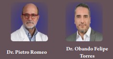
Review Article | Volume 2 | Issue 2 | JRS Jul – Dec 2022 | Page 07-12 | Pietro Romeo , Obando Felipe Torres , Federica di Pardo , Thomas Graus
DOI: 10.13107/jrs.2022.v02.i02.53

Author: Pietro Romeo [1], Obando Felipe Torres [2], Federica di Pardo [1], Thomas Graus [3]

[1] Periso Academy, Lugano, Switzerland,
[2] Cell Regeneration Medical Organization-University El Bosque, Bogotá, Colombia,

[3] Periso Medical Division, Pazzallo, Switzerland.

Address of Correspondence
Dr. Pietro Romeo, MD
Periso Academy, Lugano, Switzerland.
E-mail: romeo.p@libero.it


Abstract

Magnetism includes the tendency of the matter to react to an incoming Magnetic Field both in attractive and repulsive way. With regard to the latter phenomenon , Diamagnetism, the ultra-structure of the matter shows unpaired electrons with an antiparallel spin, the high polarity, and the absence of a proper magnetic moment. All this results in a repulsive effect which induces the movement of diamagnetic liquids and molecules, the water first, so realizing the so called diamagnetic effect . This effect involves both the extracellular and intracellular environment, with the possibility to move various diamagnetic molecules and the flux of ions across the cell membrane, acting on the metabolic processes of the biological matter. The full realization of this phenomenon requires a high intensity of the magnetic field together with the possibility to modulate two key parameters such the Frequency and the Amplitude of the impulse. This review analyses the real and possible applications of Diamagnetism in clinical practice.

Keywords: Diamagnetism, Repulsive Effect, High Intensity Magnetic Fields.


References:

1. Zablotskii V, Polyakova T, Dejneka A. Modulation of the cell membrane potential, and intracellular protein transport by high magnetic fields.Bioelectromagnetics 2021;42:27-36.
2. Hall LT, Hill CD, Cole JH, Stadler B, Caruso F, Mulvaney P, et al. Monitoring ion channel function in real-time through quantum decoherence. Proc Natl Acad Sci U S A2010;107:18777-82.
3. Viganò M, Sansone V, d’Agostino MC, Romeo P, Orfei CP, De Girolamo L. Mesenchymal stem cells as a therapeutic target of biophysical
stimulation for the treatment of musculoskeletal disorders. J Orthop Surg Res 2016;11:163.
4. Yasuda I. Piezoelectricity of living bone. J Kyoto Pref Univ Med 1953;53:325-8. Yasuda . The classic: Fundamental aspects of the treatment of bone fractures treatment by Iwao Yasuda, reprinted from J. Kyoto Med. Soc., 4:395-406,1953. Clin Orthop Relat Res 1977;124:5-8.
5. Wade B. A review of pulsed electromagnetic field (PEMF) mechanisms at a cellular level: Arationale for clinical use. Am J Health Res 2013;1:51.
6. Obando FT, Romeo P, Vergara D, di Pardo F, Soto A. The effects of low-frequency high-intensity pulsed electromagnetic fields (diamagnetic therapy) in the treatment of rare diseases: A case series preliminary study. J Neurol Exp Neural Sci 2022;4:145.
7. Roberti R, Marcianò R, Casarella A, Rania V, Palleria C, Muraca L, et al. High-intensity, low-frequency pulsed electromagnetic field as an odd treatment in a patient with mixed foot ulcer: A case report. Reports 2022;5:3.
8. National Research Council. Opportunities in High Magnetic Field Science. Washington, DC: The National Academies Press; 2005.
9. Kohout M, Savine A. Atomic shell structure and electron numbers. Int J Quantum Chem 1996;60:875-82.
10. Petrescu N, Petrescu FI. Permanent magnetic fluids. Am J Eng Appl Sci 2019;12:402-12.
11. National Council of Educational Research and Training. Website-India-Physics Textbook-Part 1-Magnetism and Matter. Ch. 5. New Delhi: National Council of Educational Research and Training; 2020. Available from:( https://www.ncert.nic.in textbook).
12. Jackson R. John Tyndall and the early history of diamagnetism. Ann Sci 2015;72:435-89.
13. Purnell MC, Skrinjar TJ. The dielectrophoretic disassociation of chloride ions and the influence on diamagnetic anisotropy in cell membranes. Discov Med 2016;22:257-73.
14. Premi E, Benussi A, La Gatta A, Visconti S, Costa A, Gilberti N, et al. Modulation of long-term potentiation-like cortical plasticity in the healthy brain with low frequency-pulsed electromagnetic fields. BMC Neurosci 2018;19:34.
15. Edmonds DT. Larmor precession as a mechanism for the detection of static and alternating magnetic fields. Bioelectrochem Bioenerg 1993;30:3-12.
16. Barnes FG, Greenebaum B. Handbook of Biological Effects of Electromagnetic Fields. 3rd ed. Florida: CRC Press; 2006.
17. World Health Organization. Magnetic Field Environmental Health Criteria 69 Magnetic Fields (EHC 69). Geneva: World Health Organization; 1987.
18. Blank M, Goodman R. A mechanism for stimulation of biosynthesis by electromagnetic fields: Charge transfer in DNAand base-pair separation. J Cell Physiol 2008;214:20-6.
19. Zablotskii V, Polyakova T, Dejneka A. Effects of high magnetic fields on the diffusion of biologically active molecules. Cells 2021;11:81.
20. Qian AR, Gao X, Zhang W, Li JB, Wang Y, Di SM, et al. Large gradient high magnetic fields affect osteoblast ultrastructure and function by disrupting collagen I or fibronectin/ab1 integrin. PLoS One 2013;8:e51036.
21. Wang Y, Chen ZH, Yin C, Ma JH, Li DJ, Zhao F, et al. Gene chip expression profiling reveals the alterations of energy metabolism related genes in osteocytes under large gradient high magnetic fields. PLoS One 2015;10:e0116359.
22. Zablotskii V, Polyakova T, Lunov O, Dejneka A. How a high-gradient magnetic field could affect cell life. Sci Rep 2016;6:37407.
23. Carnovali M, Stefanetti N, Galluzzo A, Romeo P, Mariotti M, V. Sansone. High-intensity low-frequency pulsed electromagnetic fields treatment stimulates fin regeneration in adult zebrafish-a preliminary report. Appl Sci 2022;12:7768.
24. Lin HY, Lin YJ. In vitro effects of low-frequency electromagnetic fields on osteoblast proliferation and maturation in an inflammatory environment. Bioelectromagnetics 2011;32:552-60.
25. Morris CE, Skalak TC. Acute exposure to a moderate strength static magnetic field reduces edema formation in rats. Am J Physiol Heart Circ Physiol 2008;294:H50-7.
26. Izzo M, Napolitano L, Coscia V, La Gatta A, Mariani F, Gasbarro V. The role of the diamagnetic pump (CTU Mega 18) in the physical treatment of limbs lymphoedema. A clinical study. Eur J Lymphol Rel Probl 2010;21:24-9.
27. Obando FT, Velasco JM, Soto A, Di Pardo F. Effects of High-intensity Pulsed Electromagnetic Fields (HI-PEMF) in Interstitial Lung Fibrosis due to the Anti-Synthetase Syndrome Associated with Sjogren’s Syndrome. A Case Report; 2020. p. 6.
28. Obando AF, Romeo P, Visconti S. Experience in the Respiratory Rehabilitation Program for the Treatment of Lung Interstitial Disease in Post-COVID-19 Pneumonia by Associating Low-frequency-High Intensity-Pulsed Electromagnetic Fields (Diamagnetotherapy): A Case Series Study; 2020. p. 8.
29. Obando AF, Velasco JM, Romeo P. Variable low frequency-high intensity-pulsed electromagnetic fields in the treatment of low back pain: A case series report and a review of the literature. J Orthop Res Ther 2020;5:1174.
30. Roberti R, Marcianò G, Casarella A, Rania V, Palleria C, Vocca C, et al. Diamagnetic therapy in a patient with complex regional pain syndrome Type I and multiple drug intolerance: Acase report. Reports 2022;5:18.
31. Baronio M, Sadia H, Paolacci S, Prestamburgo D, Miotti D, Guardamagna VA, et al. Molecular aspects of regional pain syndrome. Pain Res Manag 2020;2020:7697214.
32. Chan AK, Tang X, Mummaneni NV, Coughlin D, Liebenberg E, Ouyang A, et al. Lotz pulsed electromagnetic fields reduce acute inflammation in the injured rat-tail intervertebral disc JOR Spine 2019;2:e1069.
33. De Girolamo L, Viganò M, Galliera E, Stanco D, Setti S, Marazzi MG, et al. In vitro functional response of human tendon cells to different dosages of low frequency pulsed electromagnetic field. Knee Surg Sports Traumatol Arthrosc 2015;23:3443-53.
34. Vincenzi F, Targa M, Corciulo C, Gessi S, Merighi S, Setti S, et al. Pulsed electromagnetic fields increased the anti-inflammatory effect of A₂A and A₃ adenosine receptors in human T/C-28a2 chondrocytes and h FOB 1.19 osteoblasts. PLoS One 2013;8:e65561.

35. Diamagnetism as an Alternative or Integrating Cellular Therapy for COVID-19 in Knowledge from the Human Relevant Cell, Tissue, and Mathematics-based Methods as Key Tools for Understanding COVID-19 Dynamics, Kinetics, Symptoms, Risk Factors, and Non-conventional Treatment in the Coronavirus Pandemic and the Future. Chem World; 2021. p. 94-101.


How to Cite this article: Romeo P, Torres OF, Di Pardo F, Graus T |Medical Applications of Diamagnetism. | Journal of Regenerative Science | Jul – Dec 2022; 2(2): 07-12.

[Full Text HTML] [Full Text PDF] [XML]


Treatment of Morton’s neuroma with focused shock waves Comparison between shock waves and surgery

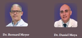
Clinical Study | Volume 2 | Issue 2 | JRS Jul – Dec 2022 | Page 13-16 | Bernard Meyer , Daniel Moya
DOI: 10.13107/jrs.2022.v02.i02.055

Author: Bernard Meyer [1], Daniel Moya [2]

[1] Orthopaedic Surgeon. Moinhos de Vento Hospital, Porto Alegre, RS, Brasil,
[2] Orthopaedic Surgeon. Hospital Británico de Buenos Aires, Argentina.

Address of Correspondence
Dr. Bernard Meyer, MD,
Orthopaedic Surgeon. Moinhos de Vento Hospital, Porto Alegre, RS, Brasil.
E-mail: bernardfabiomeyer@gmail.com


Abstract

Various treatments have been described for Morton’s neuroma. We compare the results of shock wave treatment with surgical neurectomy in a prospective series of 32 cases randomly assigned. In the group of patients treated with focused waves (17 cases), the percentage of good results was 70.6%, while in the operated patients (15 cases) it amounted to 93.2%. Poor results were found in 29.4% in shockwave group and 6.8 % in surgical group. Focused shock waves have a high, but lower success rate than surgery in the treatment of Morton’s neuromas. Nevertheless, due to their non-invasiveness and low chance of complications, they can be considered an option prior to minimally invasive and surgical techniques.

Keywords: Morton neuroma, shockwaves, neurectomy.


References:

1. Bhatia M, Thomson L. Morton’s neuroma – Current concepts review. J Clin Orthop Trauma. 2020 May-Jun;11(3):406-409. doi: 10.1016/j.jcot.2020.03.024. Epub 2020 Apr 10. PMID: 32405199; PMCID: PMC7211826.
2. Bennett G.L., Graham C.E., Mauldin D.M. Morton’s interdigital neuroma: a comprehensive treatment protocol. Foot Ankle Int. 1995;16(12):760–763. doi: 10.1177/107110079501601204.
3. Valisena S, Petri GJ, Ferrero A. Treatment of Morton’s neuroma: A systematic review. Foot Ankle Surg. 2018 Aug;24(4):271-281. doi: 10.1016/j.fas.2017.03.010. Epub 2017 Apr 5. PMID: 29409240.
4. Matthews BG, Hurn SE, Harding MP, Henry RA, Ware RS. The effectiveness of non-surgical interventions for common plantar digital compressive neuropathy (Morton’s neuroma): a systematic review and meta-analysis. J Foot Ankle Res. 2019 Feb 13;12:12. doi: 10.1186/s13047-019-0320-7. PMID: 30809275; PMCID: PMC6375221.
5. Thomson L, Aujla RS, Divall P, Bhatia M. Non-surgical treatments for Morton’s neuroma: A systematic review. Foot Ankle Surg. 2020 Oct;26(7):736-743. doi: 10.1016/j.fas.2019.09.009. Epub 2019 Nov 2. PMID: 31718949.
6. Markovic M, Crichton K, Read JW, Lam P, Slater HK. Effectiveness of Ultrasound-Guided Corticosteroid Injection in the Treatment of Morton’s Neuroma. Foot & Ankle International. 2008;29(5):483-487. doi:10.3113/FAI-2008-0483.
7. Lizano-Díez X, Ginés-Cespedosa A, Alentorn-Geli E, Pérez-Prieto D, González-Lucena G, Gamba C. et al.: Corticosteroid injection for the treatment of Morton’s neuroma: a prospective, double-blinded, randomized, placebo-controlled trial. Foot Ankle Int 2017; 38: 944–951.
8. Klontzas ME, Koltsakis E, Kakkos GA, Karantanas AH. Ultrasound-guided treatment of Morton’s neuroma. J Ultrason. 2021 Jun 7;21(85):e134-e138. doi: 10.15557/JoU.2021.0022. Epub 2021 Jun 18. PMID: 34258038; PMCID: PMC8264811.
9. Sofka CM, Adler RS, Ciavarra GA, Pavlov H. Ultrasound-guided interdigital neuroma injections: short-term clinical outcomes after a single percutaneous injection–preliminary results. HSS J. 2007 Feb;3(1):44-9. doi: 10.1007/s11420-006-9029-9. PMID: 18751769; PMCID: PMC2504098.
10. Diebold PF, Delagoutte JP. La neurolyse vraie dans le traitement du névrome de Morton [True neurolysis in the treatment of Morton’s neuroma]. Acta Orthop Belg. 1989;55(3):467-71. French. PMID: 2603689.
11. Fanucci E, Masala S, Fabiano S, Perugia D, Squillaci E, Varrucciu V, Simonetti G. Treatment of intermetatarsal Morton’s neuroma with alcohol injection under US guide: 10-month follow-up. Eur Radiol. 2004 Mar;14(3):514-8. doi: 10.1007/s00330-003-2057-7. Epub 2003 Oct 3. PMID: 14531002.
12. Gurdezi S., White T., Ramesh P. Alcohol injection for Morton’s neuroma: a five-year follow-up. Foot Ankle Int. 2013 doi: 10.1177/1071100713489555.
13. Deniz S, Purtuloglu T, Tekindur S, Cansız KH, Yetim M, Kılıckaya O, Senkal S, Bilgic S, Atim A, Kurt E. Ultrasound-guided pulsed radio frequency treatment in Morton’s neuroma. J Am Podiatr Med Assoc. 2015 Jul;105(4):302-6. doi: 10.7547/13-128.1. Epub 2015 May 6. PMID: 25945935.
14. Shah R, Ahmad M, Hanu-Cernat D, Choudhary S. Ultrasound-guided radiofrequency ablation for treatment of Morton’s neuroma: initial experience. Clin Radiol. 2019 Oct;74(10):815.e9-815.e13. doi: 10.1016/j.crad.2019.07.002. Epub 2019 Aug 10. PMID: 31409448.
15. Lee K, Hwang IY, Ryu CH, Lee JW, Kang SW. Ultrasound-Guided Hyaluronic Acid Injection for the Management of Morton’s Neuroma. Foot Ankle Int. 2018 Feb;39(2):201-204. doi: 10.1177/1071100717739578. Epub 2017 Nov 20. PMID: 29153007.
16. Pace A, Scammell B, Dhar S. The outcome of Morton’s neurectomy in the treatment of metatarsalgia. Int Orthop. 2010 Apr;34(4):511-5. doi: 10.1007/s00264-009-0812-3. Epub 2009 May 30. PMID: 19484237; PMCID: PMC2903131.
17. Kasparek M, Schneider W. Surgical treatment of Morton’s neuroma: clinical results after open excision. Int Orthop. 2013 Sep;37(9):1857-61. doi: 10.1007/s00264-013-2002-6. Epub 2013 Jul 13. PMID: 23851648; PMCID: PMC3764278.
18. Xu W, Zhang N, Li Z, Wang Y, Li X, Wang Y, Si H, Hu Y. Plantar and dorsal approaches for excision of morton’s neuroma: a comparison study. BMC Musculoskelet Disord. 2022 Oct 6;23(1):898. doi: 10.1186/s12891-022-05858-w. PMID: 36203146; PMCID: PMC9535891.
19. Akermark C, Saartok T, Zuber Z. Aprospective 2-year follow-up study of plantar incisions in the treatment of primary intermetatarsal
neuromas (Morton’s neuroma) Foot Ankle Surg. 2008;14:67–73. doi: 10.1016/j.fas.2007.10.004.
20. Akermark C, Crone H, Skoog A, Weidenhielm L. A prospective randomized controlled trial of plantar versus dorsal incisions for operative treatment of primary Morton’s neuroma. Foot Ankle Int. 2013 Sep;34(9):1198-204. doi: 10.1177/1071100713484300. Epub 2013 Apr 5. PMID: 23564425.
21. Sato G, Ferreira GF, Sevilla D, Oliveira CN, Lewis TL, Dinato MCME, Pereira Filho MV. Treatment of Morton’s neuroma with minimally invasive distal metatarsal metaphyseal osteotomy (DMMO) and percutaneous release of the deep transverse metatarsal ligament (DTML): a case series with minimum two-year follow-up. Int Orthop. 2022 Dec;46(12):2829-2835. doi: 10.1007/s00264-022-05557-0. Epub 2022 Aug 29. PMID: 36031662.
22. Bauer T, Gaumetou E, Klouche S, Hardy P, Maffulli N. Metatarsalgia and Morton’s Disease: Comparison of Outcomes Between Open Procedure and Neurectomy Versus Percutaneous Metatarsal Osteotomies and Ligament Release With a Minimum of 2 Years of Follow-Up. J Foot Ankle Surg. 2015 May-Jun;54(3):373-7. doi: 10.1053/j.jfas.2014.08.009. Epub 2014 Dec 4. PMID: 25481724.
23. Moya D, Ramón S, Schaden W, Wang CJ, Guiloff L, Cheng JH. The Role of Extracorporeal Shockwave Treatment in Musculoskeletal Disorders. J Bone Joint Surg Am. 2018 Feb 7;100(3):251-263. doi: 10.2106/JBJS.17.00661. PMID: 29406349.
24. Seok H, Kim SH, Lee SY, Park SW. Extracorporeal Shockwave Therapy in Patients with Morton’s Neuroma A Randomized, Placebo-Controlled Trial. J Am Podiatr Med Assoc. 2016 Mar;106(2):93-9. doi: 10.7547/14-131. PMID: 27031544.
25. Fridman R, Cain JD, Weil L Jr. Extracorporeal shockwave therapy for interdigital neuroma: a randomized, placebo-controlled, double-blind trial. J Am Podiatr Med Assoc. 2009 May-Jun;99(3):191-3. doi: 10.7547/0980191. PMID: 19448168.
26. Johnson, JE; Johnson, KA; Unni KK: Persistent pain after excision of interdigital neuroma. J. Bone Joint Surg. 70A:651 – 657,1988.
27. Biz C, Bonvicini B, Sciarretta G, Pendin M, Cecchetto G, Ruggieri P. Digital Ischemia after Ultrasound-Guided Alcohol Injection for Morton’s Syndrome: Case Report and Review of the Literature. J Clin Med. 2022 Oct 24;11(21):6263. doi: 10.3390/jcm11216263. PMID: 36362491; PMCID: PMC9657702.
28. Interventional procedure overview of radiofrequency ablation for symptomatic interdigital (Morton’s) neuroma. National institute for health and care excellence (NICE). https://www.nice.org.uk/guidance/ipg539/documents/radiofreque ncy-ablation-for-symptomatic-interdigital-mortons-neuromaoverview2#:~:text=ablation%20for%20symptomatic%20interdigital%20(Morton’s)%20neurom a,Interdigital%20(Morton’s)%20neuroma&text=In%20this%20procedure%2C%20a%20thin,nerve%20with%20radiofrequency%20heat20energy. Last accessed July 2022.
29. Womack JW, Richardson DR, Murphy GA, Richardson EG, Ishikawa SN. Long-term evaluation of interdigital Neuroma treated by surgical excision. Foot Ankle Int. 2008;29:574–577. doi: 10.3113/FAI.2008.0574.
30. Lorbach O, Kusma M, Pape D, Kohn D, Dienst M. Influence of deposit stage and failed ESWT on the surgical results of arthroscopic treatment of calcifying tendonitis of the shoulder, Knee Surg. Sports Traumatol. Arthrosc. 16 (5) (2008) 516e-521, http://dx.doi.org/10.1007/s00167-008-0507-0.


How to Cite this article: Meyer B, Moya D | Treatment of Morton’s neuroma with focused shock waves Comparison between shock waves and surgery. | Journal of Regenerative Science | Jul – Dec 2022; 2(2): 13-16.

[Abstract Text HTML] [Full Text PDF] [XML]


Use of shock waves in dental medicine

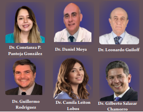
Literature Review | Volume 2 | Issue 2 | JRS Jul – Dec 2022 | Page 17-20 | Constanza P. Pantoja González, Daniel Moya, Leonardo Guiloff, Guillermo Rodríguez, Camila Leiton Lobos, Gilberto Salazar Chamorro
DOI: 10.13107/jrs.2022.v02.i02.57

Author: Constanza P. Pantoja González [1], Daniel Moya [2], Leonardo Guiloff [3], Guillermo Rodríguez [4], Camila Leiton Lobos [5], Gilberto Salazar Chamorro [6]

[1] Dental Surgeon, Diego Portales University, Chile.
[2] Department of Orthopaedics, Hospital Británico de Buenos Aires, Argentina,

[3] Surgeon, Specialist in Traumatology and Orthopedics, University of Chile, Founding Partner and Past President of ACHITOC and ONLAT, Chile,
[4] Dental Surgeon, University of Buenos Aires; Endodontics Specialist, Maimonides University, Bs As; , Specialist in Oral Implantology, Catholic University of Argentina, Buenos Aires.

[5] Dental Surgeon, Diego Portales University, Specialist in Oral Maxillofacial Implantology Andrés Bello University, Chile,
[6] Dental Surgeon, Pontifical Javierana University, Colombia. Specialist in Oral Maxillofacial Implantology, Professor at San Sebastián University, Chile.

Address of Correspondence
Dr. Constanza P. Pantoja Gonzalez, DDS,

Dental Surgeon, Diego Portales University, Chile.
E-mail: coni.panto@gmail.com


References:

1. Moya D, Ramón S, Schaden W, Wang CJ, Guiloff L, Cheng JH. The Role of Extracorporeal Shockwave Treatment in Musculoskeletal Disorders. J Bone Joint Surg Am. 2018 Feb 7;100(3):251-263. doi: 10.2106/JBJS.17.00661. PMID: 29406349.
2. Olivares A, Schuh CMAP, Aguayo S. Extracorporeal Shockwave Treatment for Managing Biofilm-mediated Infections in Dentistry: The Current Knowledge and Future Perspectives. Journal of Regenerative Science. Jan – Jun 2022; 2(1): 22-26.
3. Alshihri A. Translational Applications of Extracorporeal Shock Waves in Dental Medicine: A Literature Review. Biomedicines. 2022;10(4):902. https://doi.org/10.3390/biomedicines10040902
4. Amengual-Penafiel L, Jara-Sepúlveda MC, Parada-Pozas L, Marchesani-Carrasco F, Cartes-Velásquez R, Galdames-Gutiérrez B. Immunomodulation of Osseointegration Through Extracorporeal Shock Wave Therapy. Dent Hypotheses 2018;9:45-50.
5. Goyal, Eva, et al. “Extra Corporeal Shock Wave – A New Wave of Therapy.” Dental Journal of Advance Studies. 2015; 3 (3): 129–34, https://doi.org/10.1055/s-0038-1672027.
6. Li X, Chen M, Li L, Qing H, Zhu Z. Extracorporeal shock wave therapy: a potential adjuvant treatment for peri-implantitis. Med Hypotheses. 2010 Jan;74(1):120-2. doi: 10.1016/j.mehy.2009.07.025. Epub 2009 Aug 8. PMID: 19666209.
7. Morandi P, Corbella S, Cavalli N, Francetti L. Applicazioni delle onde d’urto in odontoiatria: revisione narrativa. Dental Cadmos. Oct 2019; 87(8). doi: 10.19256/d.cadmos.08.2019.04
8. Show S, Kumar Giri P, Debnath T, Ashit Kumar Pal A. Extracorporeal shockwave therapy… “unveiling new horizons in periodontology”- an overview. J Indian Dental Assoc. 2020; 36 (1): 44-47.
9. Song WP, Ma XH, Sun YX, Zhang L, Yao Y, Hao XY, Zeng YJ. Extracorporeal shock wave therapy (ESWT) may be helpful in the osseointegration of dental implants: A hypothesis. Medical Hypotheses. 2020; 145. https://doi.org/10.1016/j.mehy.2020.110294
10. Venkatesh Prabhuji ML, Khaleelahmed S, Vasudevalu S, Vinodhini K. Extracorporeal shock wave therapy in periodontics: Anew paradigm. J Indian Soc Periodontol. 2014 May;18(3):412-5. doi: 10.4103/0972-124X.134597. PMID: 25024562; PMCID: PMC4095641.
11. Goker F, Sansone V, Applefield RC, Taschieri S, Del Fabbro M. Clinical applications of shock waves in dentistry. J Biol Regul Homeost Agents. 2019 September-October;33(5):1591-1595. doi: 10.23812/19-15L. PMID: 31565915.
12. Özkan E, Özkan TH. Effects of Extracorporeal Shock Wave Therapy in The Maxillofacial Surgery Practice – A Systematic Review. International Journal of Human and Health Sciences. 2019; 3 (4): 186-195.
13. Elisetti N. Extracorporeal shock wave therapy (ESWT): An emerging treatment for peri-implantitis. Med Hypotheses. 2021 May;150:110565. doi: 10.1016/j.mehy.2021.110565. Epub 2021 Mar 23. PMID: 33799162.
14. Cai Z, Falkensammer F, Andrukhov O, Chen J, Mittermayr R, Rausch-Fan X. Effects of Shock Waves on Expression of IL-6, IL-8, MCP-1, and TNF-α Expression by Human Periodontal Ligament Fibroblasts: An In Vitro Study. Med Sci Monit. 2016 Mar 20;22:914-21. doi: 10.12659/msm.897507. PMID: 26994898; PMCID: PMC4805137.
15. Novak KF, Govindaswami M, Ebersole JL, Schaden W, House N, Novak MJ. Effects of low-energy shock waves on oral bacteria. J Dent Res. 2008 Oct;87(10):928-31. doi: 10.1177/154405910808701009. PMID: 18809745.
16. Olivares A, Schuh CMAP, Aguayo S. Inhibitory effect of FhESWT on Streptococcus Mutans biofilm formation in-vitro. 2022 IADR/APR General sesión. Final Presentation ID: 0942. https://iadr.abstractarchives.com/abstract/22iags-3722065/inhibitory-effect-of-fheswt-on-streptococcus-mutans-biofilm-formation-in-vitro Last accessed August 2022.
17. Altuntaş EE, Oztemur Z, Ozer H, Müderris S. Effect of extracorporeal shock waves on subcondylar mandibular fractures. J Craniofac Surg. 2012 Nov;23(6):1645-8.doi: 10.1097/SCS.0b013e31825e38a2. PMID: 23147295.
18. Atsawasuwan P, Chen Y, Ganjawalla K, Kelling AL, Evans CA. Extracorporeal shockwave treatment impedes tooth movement in rats. Head Face Med. 2018 Nov 12;14(1):24. doi: 10.1186/s13005-018-0181-5. PMID: 30419912; PMCID: PMC6233511.
19. Chen Y, Ganjawalla K, Oubaidin M, Kelling A, Evans C, Atsawasuwan P. Effect of Shockwave Therapy on Orthodontic Tooth Movement. 2015 IADR/AADR/CADR General Session (Boston, Massachusetts). Final Presentation ID: 3987https://iadr.abstractarchi ves.com/abstract/15iags-2104243/effect-of-shockwave-therapy-on-orthodontic-tooth-movement Last accessed August,2022.
20. Datey A, Thaha CSA, Patil SR, Gopalan J, Chakravortty D. Shockwave Therapy Efficiently Cures Multispecies Chronic Periodontitis in a Humanized Rat Model. Front Bioeng Biotechnol. 2019 Dec 13;7:382. doi: 10.3389/fbioe.2019.00382. PMID: 31911896; PMCID: PMC6923175.
21. Ginini JG, Maor G, Emodi O, Shilo D, Gabet Y, Aizenbud D, Rachmiel A. Effects of Extracorporeal Shock Wave Therapy on Distraction Osteogenesis in Rat Mandible. Plast Reconstr Surg. 2018 Dec;142(6):1501-1509. doi: 10.1097/PRS.0000000000004980. Erratum in: Plast Reconstr Surg. 2019 Feb;143(2):654. PMID: 30188470.
22. Göl EB, Özkan N, Bereket C, Önger ME. Extracorporeal Shock-Wave Therapy or Low-Level Laser Therapy: Which is More Effective in Bone Healing in Bisphosphonate Treatment? J Craniofac Surg. 2020 Oct;31(7):2043-2048. doi: 10.1097/SCS.0000000000006506. PMID:32371691.
23. Hazan-Molina H, Kaufman H, Reznick ZA, Aizenbud D. [Orthodontic tooth movement under extracorporeal shock wave therapy: the characteristics of the inflammatory reaction–a preliminary study]. Refuat Hapeh Vehashinayim (1993). 2011 Jul;28(3):55-60, 71. Hebrew. PMID: 21939106.
24. Hazan-Molina H, Kaufman H, Reznick ZA, Aizenbud D. Cytokine Concentration During Orthodontic Tooth Movement Under Shock Wave Therapy. 2012 Pan European Region Meeting (Helsinki, Finland). Poster session. https://iadr.abstractarchives.com/abstract/per12-167553/cytokine-concentration-during-orthodontic-tooth-movement-under-shock-wave-therapy Last accessed August 20,2022.
25. Hazan-Molina H, Reznick ZA, Kaufman H, Aizenbud D. Assessment of IL-1β and VEGF concentration in a rat model during orthodontic tooth movement and extracorporeal shock wave therapy. Archives of Oral Biology. 2013; 58 (2): 142-150.
26. Hazan-Molina H, Reznick AZ, Kaufman H, Aizenbud D. Periodontal cytokines profile under orthodontic force and extracorporeal shock wave stimuli in a rat model. J Periodontal Res. 2015 Jun;50(3):389-96. doi: 10.1111/jre.12218. Epub 2014 Jul 29. PMID: 25073624.
27. Hazan-Molina H, Aizenbud I, Kaufman H, Teich S, Aizenbud D. The Influence of Shockwave Therapy on Orthodontic Tooth Movement Induced in the Rat. Adv Exp Med Biol. 2016;878:57-65. doi: 10.1007/5584_2015_179. PMID: 26542601.
28. Hazan-Molina H, Gabet Y, Aizenbud D. Accelerated Orthodontic Tooth Movement – Fiction or Reality. 2016 IADR/PER Congress (Jerusalem, Israel) Final Presentation ID: 0070https://iadr.abstractarchives.com/abstract/per16-2530206/accelerated-orthodontic-tooth-movement–fiction-or-reality Last accessed August 2022.
29. Hazan-Molina H, Gabet Y, Aizenbud I, Aizenbud N, Aizenbud D. Orthodontic force and extracorporeal shock wave therapy: Assessment of orthodontic tooth movement and bone morphometry in a rat model. Arch Oral Biol. 2022 Feb;134:105327. doi: 10.1016/j.archoralbio.2021.105327. Epub 2021 Nov 29. PMID: 34891101.
30. Lai JP, Wang FS, Hung CM, Wang CJ, Huang CJ, Kuo YR. Extracorporeal shock wave accelerates consolidation in distraction osteogenesis of the rat mandible. J Trauma. 2010 Nov;69(5):1252-8. doi: 10.1097/TA.0b013e3181cbc7ac. PMID: 20404761.
31. Özkan E, Bereket MC, Önger ME, Polat AV. The Effect of Unfocused Extracorporeal Shock Wave Therapy on Bone Defect Healing in Diabetics. J Craniofac Surg. 2018 Jun;29(4):1081-1086. doi: 10.1097/SCS.0000000000004303. PMID: 29461364.
32. Özkan E, Bereket MC, Şenel E, Önger ME. Effect of Electrohydraulic Extracorporeal Shockwave Therapy on the Repair of Bone Defects Grafted With Particulate Allografts. J Craniofac Surg. 2019 Jun;30(4):1298-1302. doi: 10.1097/SCS.0000000000005213. PMID: 31166268.
33. Sathishkumar S, Meka A, Dawson D, House N, Schaden W, Novak MJ, Ebersole JL, Kesavalu L. Extracorporeal shock wave therapy induces alveolar bone regeneration. J Dent Res. 2008 Jul;87(7):687-91. doi: 10.1177/154405910808700703. PMID: 18573992.
34. Woodmansey K, White R, Rhodes S, Kramer P. Effects of Extracorporeal Shockwave Therapy: A Pilot Study Using a Rat Model. 2015 IADR/AADR/CADR General Session (Boston, Massachusetts). https://iadr.abstractarchives.com/abstract/15iags-2068595/effects-of-extracorporeal-shockwave-therapy-a-pilot-study-using-a-rat-model Last accessed August 2022.
35. Bereket C, Çakir-Özkan N, Önger ME, Arici S. The Effect of Different Doses of Extracorporeal Shock Waves on Experimental Model Mandibular Distraction. J Craniofac Surg. 2018 Sep;29(6):1666-1670. doi: 10.1097/SCS.0000000000004571. PMID: 29742568.
36. Demir O, Arici N. Dose-related effects of extracorporeal shock waves on orthodontic tooth movement in rabbits. Sci Rep. 2021 Feb 9;11(1):3405. doi: 10.1038/s41598-021-82997-5. PMID: 33564049; PMCID: PMC7873214.
37. Onger ME, Bereket C, Sener I, Ozkan N, Senel E, Polat AV. Is it possible to change of the duration of consolidation period in the distraction osteogenesis with the repetition of extracorporeal shock waves? Med Oral Patol Oral Cir Bucal. 2017 Mar 1;22(2):e251-e257. doi: 10.4317/medoral.21556. PMID: 28160590; PMCID: PMC5359710.
38. Senel E, Ozkan E, Bereket MC, Onger ME. The assessment of new bone formation induced by unfocused extracorporeal shock wave therapy applied on pre-surgical phase of distraction osteogenesis. Eur Oral Res. 2019 Sep;53(3):125-131. doi: 10.26650/eor.20190041. Epub 2019 Sep 1. PMID: 31579893; PMCID: PMC6761485.
39. Vares YE, Shtybel NV, Dudash AP. Does extracorporeal shock wave therapy leads to restitution of postoperative bone defects on mandible? an experimental study in rabbit model. Romanian Journal of Oral Rehabilitation. 2019; 11 (4): 234-241.
40. Ahmed EAE, Eldibany M M, Melek LF; Abdelnaby HM. Comparative study between the effect of shockwave therapy and low-intensity pulsed ultrasound (lipus) on bone healing of mandibular fractures (clinical & radiographic study). Alexandria Dental J. 2022;5,47,(1):Page 29-35.
41. Falkensammer F, Rausch-Fan X, Arnhart C, Krall C, Schaden W, Freudenthaler J Impact of extracorporeal shock-wave therapy on the stability of temporary anchorage devices in adults: A single-center, randomized, placebo-controlled clinical trial. American Journal of Orthodontics and Dentofacial Orthopedics. 2014; 146 (4):413- 422.https://doi.org/10.1016/j.ajodo.2014.06.008
42. Falkensammer F, Arnhart C, Krall C, Schaden W, Freudenthaler J, Bantleon HP. Impact of extracorporeal shock wave therapy (ESWT) on orthodontic tooth movement-a randomized clinical trial. Clin Oral Investig. 2014 Dec;18(9):2187-92. doi: 10.1007/s00784-014-1199-0. Epub 2014 Feb 19. PMID: 24549763.
43. Falkensammer F, Schaden W, Krall C, Freudenthaler J, Bantleon HP. Effect of extracorporeal shockwave therapy (ESWT) on pulpal blood flow after orthodontic treatment: a randomized clinical trial. Clin Oral Investig. 2016 Mar;20(2):373-9. doi: 10.1007/s00784-015-1525-1. Epub 2015 Jul 17. PMID: 26179985.
44. Falkensammer F, Rausch-Fan X, Schaden W, Kivaranovic D, Freudenthaler J. Impact of extracorporeal shockwave therapy on tooth mobility in adult orthodontic patients: a randomized single-center-placebo-controlled clinical trial. J Clin Periodontol. 2015 Mar;42(3):294-301. doi: 10.1111/jcpe.12373. Epub 2015 Feb 20. PMID: 25640577.
45. Pfaff JA, Boelck B, Bloch W, Nentwig GH. Growth Factors in Bone Marrow Blood of the Mandible With Application of Extracorporeal
Shock Wave Therapy. Implant Dent. 2016 Oct;25(5):606-12. doi: 10.1097/ID.0000000000000452. PMID: 27504532.
46. What is periodontitis? European Federation of Periodontology. https://www.efp.org/for-patients/what-is-periodontitis/#:~:text=Periodontitis%20is%20a%20gum%20disease, lead%20to%20other%20health%20problems. Last accessed August
2022.
47. Vestby LK, Grønseth T, Simm R, Nesse LL. Bacterial Biofilm and its Role in the Pathogenesis of Disease. Antibiotics (Basel). 2020 Feb
3;9(2):59. doi: 10.3390/antibiotics9020059. PMID: 32028684; PMCID: PMC7167820.
48. Hernández-Jiménez E, Del Campo R, Toledano V, Vallejo-Cremades MT, Muñoz A, Largo C, Arnalich F, García-Rio F, Cubillos-Zapata C, López-Collazo E. Biofilm vs. planktonic bacterial mode of growth: which do human macrophages prefer? Biochem Biophys Res Commun. 2013 Nov 29;441(4):947-52. doi: 0.1016/j.bbrc.2013.11.012. Epub 2013 Nov 14. PMID: 24239884.
49. Sharma D, Misba L, Khan AU. Antibiotics versus biofilm: an emerging battleground in microbial communities. Antimicrob Resist Infect Control. 2019 May 16;8:76. doi: 10.1186/s13756-019-0533-3. PMID: 31131107; PMCID: PMC6524306.
50. Prathapachandran J, Suresh N. Management of peri-implantitis. Dent Res J (Isfahan). 2012 Sep;9(5):516-21. doi: 10.4103/1735- 3327.104867. PMID: 23559913; PMCID: PMC3612185. 51. Astolfi V, Ríos-Carrasco B, Gil-Mur FJ, Ríos-Santos JV, Bullón B, Herrero-Climent M, Bullón P. Incidence of Peri-Implantitis and Relationship with Different Conditions: A Retrospective Study. Int J Environ Res Public Health. 2022 Mar 31;19(7):4147. doi: 10.3390/ijerph19074147. PMID: 35409826; PMCID: PMC8998347.
52. Guglielmotti MB, Olmedo DG, Cabrini RL. Research on implants and osseointegration. Periodontol 2000. 2019 Feb;79(1):178-189. doi: 10.1111/prd.12254. PMID: 30892769.
53. Ghannam MG, Alameddine H, Bordoni B. Anatomy, Head and Neck, Pulp (Tooth) [Updated 2022 Aug 8]. In: StatPearls [Internet]. Treasure Island (FL): StatPearls Publishing; 2022 Jan-. Available from: https://www.ncbi.nlm.nih.gov/books/NBK537112/ [Last accessed on Jul 2022].
54. “Orthodontics”. Britannica, The Editors of Encyclopaedia. 2018; Encyclopedia Britannica, 4 Jan. 2018, https://www.britannica.com/science/orthodontics. Last accessed August 2022].


How to Cite this article: Pantoja C, Moya D, Guiloff L, Rodríguez G, Leiton Lobos C, Salazar Chamorro G | Use of Shock Waves in Dental Medicine. | Journal of Regenerative Science | Jul – Dec 2022; 2(2): 17-20.

[Full Text HTML] [Full Text PDF] [XML]


Extracorporeal shock wave treatment in plantar fasciitis with an associated neuropathic component. How to optimize the result?

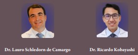
Case Report | Volume 2 | Issue 2 | JRS Jul – Dec 2022 | Page 21-23 | Lauro Schledorn de Camargo , Ricardo Kobayashi
DOI: 10.13107/jrs.2022.v02.i02.59

Author: Lauro Schledorn de Camargo [1], Ricardo Kobayashi [2]

[1] Orthopedic Surgeon at LC Clinic, Jundiai-SP Brazil, Brazil,
[2] Pain Center, Department of Neurology, University of São Paulo, Brazil.

Address of Correspondence
Dr. Lauro Schledorn de Camargo, MD,
Orthopedic Surgeon at LC Clinic, Jundiai-SP Brazil, Brazil.
E-mail: laurosch@hotmail.com


Abstract

Introduction: Current evidence supports the use of radial pressure wave and focused extracorporeal shock wave treatment (ESWT) for the treatment of chronic plantar fasciitis that does not improve with conservative treatment. Studies show that a quarter of plantar fasciitis may have an associated neuropathic component and the literature shows that neuropathic pain causes more intense pain and greater functional disability. However, there is a lack of literature on the results of ESWT in tendinopathies associated with the neuropathic pattern.
Case report: We report a case of plantar fasciitis with central sensitization and associated neuropathic component. At first, pregabalin 75mg twice a day was used, which improved the neuropathic pattern. After that, 3 sessions were performed with piezoelectric ESWT with energy of 0.12 mJ/mm2, 2000 impulses at a frequency of 8 Hz, once a week for three weeks. The patient was followed up for 3 months and had complete improvement of symptoms without functional limitation for activities of daily living.
Conclusion: This case report serves to draw attention to the importance of evaluating and treating the neuropathic pattern associated with tendinopathies in order to optimize the therapeutic result. However, randomized clinical trials are lacking to determine the real difference in results between using ESWT in nociceptive pain or in mixed pain with an associated neuropathic component.

Keywords: Chronic pain, plantar fasciitis, mixed pain, neuropathic pain, shockwaves.


References:

1. Moya D, Ramón S, Schaden W, Wang CJ, Guiloff L, Cheng JH. The Role of Extracorporeal Shockwave Treatment in Musculoskeletal Disorders. J Bone Joint Surg Am. 2018 Feb 7 ; 100 (3):251-263. doi:10.2106/JBJS.17.00661.
2. Schneider HP, Baca JM, Carpenter BB, Dayton PD, Fleischer AE, Sachs BD. American College of Foot and Ankle Surgeons Clinical Consensus Statement: Diagnosis and Treatment of Adult Acquired Infracalcaneal Heel Pain. J Foot Ankle Surg. 2018 Mar-Apr;57(2):370-381. doi: 10.1053/j.jfas.2017.10.018.
3. Wheeler PC. Neuropathic pain may be common in chronic lower limb tendinopathy: a prospective cohort study. Br J Pain. 2017 Feb;11(1):16-22. doi: 10.1177/2049463716680560.
4. Bouhassira D, Attal N, Alchaar H, Boureau F, Brochet B, Bruxelle J, Cunin G, Fermanian J, Ginies P, Grun-Overdyking A, Jafari-Schluep H, Lantéri-Minet M, Laurent B, Mick G, Serrie A, Valade D, Vicaut E. Comparison of pain syndromes associated with nervous or somatic lesions and development of a new neuropathic pain diagnostic questionnaire (Dn4). Pain. 2005 Mar;114(1-2):29-36. doi: 10.1016/j.pain.2004.12.010.
5. Santos JG, Brito JO, de Andrade DC, Kaziyama VM, Ferreira KA, Souza I, Teixeira MJ, Bouhassira D, Baptista AF. Translation to Portuguese and validation of the Douleur Neuropathique 4 questionnaire. J Pain. 2010 May;11(5):484-90. doi: 10.1016/j.jpain.2009.09.014.
6. Maki M, Ikoma K, Kido M, Hara Y, Sawada K, Ohashi S, Kubo T. Magnetic resonance imaging findings of chronic plantar fasciitis before and after extracorporeal shock wave therapy. Foot (Edinb). 2017 Dec;33:25-28. doi: 10.1016/j.foot.2017.10.002.
7. Wheeler PC. Up to a quarter of patients with certain chronic recalcitrant tendinopathies may have central sensitisation: a prospective cohort of more than 300 patients. Br J Pain. 2019 Aug;13(3):137-144. doi: 10.1177/2049463718800352.
8. Colloca L, Ludman T, Bouhassira D, Baron R, Dickenson AH, Yarnitsky D, Freeman R, Truini A, Attal N, Finnerup NB, Eccleston C, Kalso E, Bennett DL, Dworkin RH, Raja SN. Neuropathic pain. Nat Rev Dis Primers. 2017 Feb 16;3:17002. doi: 10.1038/nrdp.2017.2.
9. Attal N, Cruccu G, Baron R, Haanpää M, Hansson P, Jensen TS, Nurmikko T. EFNS guidelines on the pharmacological treatment of neuropathic pain: 2010 revision. Eur J Neurol. 2010 Sep;17(9):1113-e88. doi: 10.1111/j.1468-1331.2010.02999.x.
10. Finnerup NB, Attal N, Haroutounian S, McNicol E, Baron R, Dworkin RH, Gilron I, Haanpää M, Hansson P, Jensen TS, Kamerman PR, Lund K, Moore A, Raja SN, Rice AS, Rowbotham M, Sena E, Siddall P, Smith BH, Wallace M. Pharmacotherapy for neuropathic pain in adults: a systematic review and meta-analysis. Lancet Neurol. 2015 Feb;14(2):162-73. doi: 10.1016/S1474-4422(14)70251-0.
11. Gerdesmeyer L, Frey C, Vester J, Maier M, Weil L Jr, Weil L Sr, Russlies M, Stienstra J, Scurran B, Fedder K, Diehl P, Lohrer H, Henne M, Gollwitzer H. Radial extracorporeal shock wave therapy is safe and effective in the treatment of chronic recalcitrant plantar fasciitis: results of a confirmatory randomized placebo-controlled multicenter study. Am J Sports Med. 2008 Nov;36(11):2100-9. doi: 10.1177/0363546508324176.
12. Gollwitzer H, Saxena A, DiDomenico LA, Galli L, Bouché RT, Caminear DS, Fullem B, Vester JC, Horn C, Banke IJ, Burgkart R, Gerdesmeyer L. Clinically relevant effectiveness of focused extracorporeal shock wave therapy in the treatment of chronic plantar fasciitis: a randomized, controlled multicenter study. J Bone Joint Surg Am. 2015 May 6;97(9):701-8. doi: 10.2106/JBJS.M.01331.
13. Morrissey D, Cotchett M, Said J’Bari A, Prior T, Griffiths IB, Rathleff MS, Gulle H, Vicenzino B, Barton CJ. Management of plantar heel pain: a best practice guide informed by a systematic review, expert clinical reasoning and patient values. Br J Sports Med. 2021 Oct;55(19):1106-1118. doi: 10.1136/bjsports-2019-101970.


How to Cite this article: Camargo Lsd, Kobayashi R |Extracorporeal shock wave treatment in plantar fasciitis with an associated neuropathic component. How to optimize the result?. | Journal of Regenerative Science | Jul – Dec 2022; 2(2): 21-23.

[Full Text HTML] [Full Text PDF] [XML]