Shock Waves and Radial Pressure Waves: Time to Put a Clear Nomenclature into Practice

Review Article | Volume 1 | Issue 1 | JRS December 2021 | Page 4-8 | Achim M. Loske, Daniel Moya DOI: 10.13107/jrs.2021.v01.i01.005

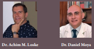
Author: Achim M. Loske [1], Daniel Moya [2]

[1] Centro de Física Aplicada y Tecnología Avanzada, Universidad Nacional Autónoma de México, Blvd. Juriquilla 3001, Querétaro, Qro., 76230, México.

[2] Department of Orthopaedic, Servicio de Ortopedia y Traumatología, Hospital Británico de Buenos Aires.

Address of Correspondence:
Dr. Daniel Moya, MD
Department of Orthopaedic, Servicio de Ortopedia y Traumatología, Hospital Británico de Buenos Aires.
E-mail: drdanielmoya@yahoo.com.ar


Abstract

Extracorporeal focused shock wave therapy and radial pressure wave therapy are noninvasive approaches with high success rates that hold promise for treating a rapidly increasing number of clinical indications. However, reports, presentations at scientific meetings, and information published by manufacturers reflect confusion in the terminology used. This situation is worrisome because both desired and undesired biological effects depend on the pressure profile and the physical parameters used. Moreover, in many cases, the detailed biological mechanisms involved are yet not fully understood. Only a clear knowledge of the physical concepts can enable comparison among and improvement of treatment protocols and technology. Fortunately, specific definitions and recommendations have been agreed upon by scientific societies promoting international standardization. The main goal of this article is to raise awareness of the importance of having a clear nomenclature worldwide and explain some of the concepts based on the international consensus that has been accepted to date.

Keywords: Shock waves, Radial pressure waves, Physical parameters .


Reference:

  1. Britannica, the Editors of Encyclopaedia. International System of Units. Encyclopedia Britannica, 30 Jul; 2020. Available from: https://www.britannica.com/science/International-System-of-Units [Last accessed on 2021 May 8].
  2. Metric Convention of 1875. US Metric Association. Available from: https://usma.org/laws-and-bills/metric-convention-of-1875 [Last accessed on 2021 May 8].
  3. Delius M, Brendel W. Historical roots of lithotripsy. J Lithotr Stone Dis 1990;2:161-3.
  4. Chaussy C, Eisenberger F, Forssmann B. Epochs in endourology; extracorporeal shockwave lithotripsy (ESWL): A chronology. J Endourol 2007;21:1249-53.
  5. Loske AM. Medical and Biomedical Applications of Shock Waves. Cham, Switzerland: Springer International; 2017. p. 19-42.
  6. Türk C, Knoll T, Petrik A, Sarica K, Skolarikos A, Straub M, et al. Guidelines on Urolithiasis. Arnhem, Netherlands: European Association of Urology; 2015. Available from: https://uroweb.org/wp-content/uploads/22-Urolithiasis_LR_full.pdf [Last accessed on 2021 May 8].
  7. Graff J, Richter KD, Pastor J. Effect of high energy shock waves on bony tissue. Urol Res 1988;16:252-8.
  8. Karpmann RR, Magee FP, Gruen TWS, Mobley T. The lithotriptor and its potential use in the revision of total hip arthroplasty. Orthop Rev 1987;16:38-42.
  9. Valchanou VD, Michailov P. High energy shock waves in the treatment of delayed and nonunion of fractures. Int Orthop 1991;15:181-4.
  10. Rompe JD, Rumler F, Hopf C, Nafe B, Heine J. Extracorporal shock wave therapy for calcifying tendinitis of the shoulder. Clin Orthop Relat Res 1995;321:196-201.
  11. Haupt G. Use of extracorporeal shock waves in the treatment of pseudarthrosis, tendinopathy and other orthopedic diseases. J Urol 1997;158:4-11.
  12. Lohrer H, Gerdesmeyer L, editors. Shock wave therapy in practice. In: Multidisciplinary Medical Applications. Heilbronn: Buchverlag; 2014. p. 50-69.
  13. Speed C. A systematic review of shockwave therapies in soft tissue conditions: Focusing on the evidence. Br J Sports Med 2014;48:1538-42.
  14. Mittermayr R, Antonic V, Hartinger J, Kaufmann H, Redl H, Téot L, et al. Extracorporeal shock wave therapy (ESWT) for wound healing: Technology, mechanisms, and clinical efficacy. Wound Repair Regen 2012;20:456-65..
  15. Moya D, Ramón S, Schaden W, Wang CJ, Guiloff L, Cheng JH. The role of extracorporeal shockwave treatment in musculoskeletal disorders. J Bone Joint Surg Am 2018;100:251-63.
  16. Auersperg V. DIGEST Guidelines for Extracorporeal Shock Wave Therapy. Physics and Technology of ESWT. Available from: http://www.setoc.es/docs/DIGEST%20guidelines_June%202019_E_A.pdf [Last accessed on 2021 May 8].
  17. IEC 61846 International Standard Ultrasonics/Pressure Pulse Lithotripters/Characteristics of Fields. Vol. 18. Geneva, Switzerland: International Electrotechnical Commission; 1998.
  18. Ueberle F, Rad AJ. Ballistic pain therapy devices: Measurement of pressure pulse parameters. Biomed Tech 2012;57:700-3.
  19. International Society for Medical Shockwave Therapy: Physical principles of ESWT Basic Physical Principles. Available from: https://www.shockwavetherapy.org/about-eswt/physical-principles-of-eswt [Last accessed on 2021 May 8].
  20. Novak P. Physics: F-SW and R-SW. Shock wave therapy in practice. Basic information on focused and radial shock wave physics. In: Lohrer H, Gerdesmeyer L, editors. Multidisciplinary Medical Applications. Heilbronn: Buchverlag; 2014. p. 28-49.
  21. Federación Ibero-Latinoamericana de Ondas de Choque e Ingeniería Tisular (Onlat) Ondas de Choque en Medicina: La Nueva Frontera. Available from: https://onlat.net/?page_id=2491 [Last accessed on 2021 May 8].
  22. European Commission DG Health and Consumer. Medical Devices: Guidance Document. Classification of medical devices. MEDDEV 2. 4/1 Rev. 9 June 2010. Available from: https://pdf4pro.com/download/medical-devices-guidance-document-6fa97.html [Last accessed on 2021 May 8].
  23. Eid J. ISMST Consensus Statement Terms and Definitions. Available from: https://www.shockwavetherapy.org/fileadmin/user_upload/dokumente/PDFs/Formulare/Consensus_MBRadial_pressure_wave_2017_SS.pdf [Last accessed on 2021 May 8].
  24. Consenso de la Federación Ibero-Latinoamericana de Ondas de Choque e Ingeniería Tisular Sobre las Bases Físicas de las Ondas de Choque Focales y de las Ondas de Presión Radial. Available from: https://onlat.net/?page_id=2497 [Last accessed on 2021 May 8].
  25. Consensus Statement on ESWT Indications and Contraindications. Available from: https://www.shockwavetherapy.org/fileadmin/user_upload/dokumente/PDFs/Formulare/ISMST_consensus_statement_on_indications_and_contraindications_20161012_final.pdf [Last accessed on 2021 May 8].
  26. Recommendation Statement of the Conjoint Physics Working Group of ISMST and DIGEST on ESWT study design and publication. ISMST Recommendations. Available from: https://www.shockwavetherapy.org/about-eswt/ismst-recommendations [Last accessed on 2021 May 8].
  27. Ramon S, Español A, Yebra M, Morillas JM, Unzurrunzaga R, Freitag K, et al. Ondas de choque. Evidencias y recomendaciones SETOC (Sociedad Española de Tratamientos con Ondas de Choque). Rehabilitación (Madr) 2021;55:291-300.
  28. Sociedade Médica Brasileira de Tratamento por Ondas de Choque. Physical Features. Available from: https://www.sbtoc.org.br/aspectos-fisicos [Last accessed on 2021 May 8].
  29. Deutschsprachige Internationale Gesellschaft für Extrakorporale Stoßwellentherapie: Technology, Technical Differences. Available from: https://www.digest-ev.de/gesellschaft/geschichte.html [Last accessed on 2021 May 8].


How to Cite this article: Loske AM, Moya D | Shock waves and radial pressure waves: time to put a clear nomenclature into practice. | Journal of Regenerative Science | December 2021; 1(1): 4-8.

[Full Text HTML] [Full Text PDF] [XML]


Case Report: Focused Shock Waves as a Treatment Option in Failed Rotator Cuff Calcification Surgery

Case Report | Volume 1 | Issue 1 | JRS December 2021 | Page 51-54 | Daniel Moya. DOI: 10.13107/jrs.2021.v01.i01.027

Author: Daniel Moya [1]

[1] Department of Orthopaedic, Servicio de Ortopedia y Traumatología, Hospital Británico de Buenos Aires, Argentina.

Address of Correspondence
Dr. Daniel Moya, MD
Department of Orthopaedic, Servicio de Ortopedia y Traumatología, Hospital Británico de Buenos Aires, Argentina.
E-mail: drdanielmoya@yahoo.com.ar


Abstract

Introduction: Focused extracorporeal shock wave treatment has emerged as an alternative therapy before invasive procedures when conservative treatment has failed in rotator cuff calcifications. It can also be used when surgery has failed.
Case Report: We report a case of failed surgery in which focused shock waves were used for treatment. We applied three sessions of focused electromagnetic waves (Dornier Compact Alpha) with an energy level of 0.20 mJ/mm2, 2000 pulses per sesión, every 2 weeks. The pain gradually disappeared and mobility was regaining. The radiographic control 2 months after the last session showed the total disappearance of the calcification. The end result was the complete disappearance of the symptoms.
Conclusion: The effectiveness of shock waves, their non-invasiveness, safety, and cost efficiency justify their use both as an option before an invasive technique and when surgery has failed in the treatment of rotator cuff calcifications.
Keywords: Rotator cuff calcifications, Shock waves, Failed surgery.


References:

1. Bosworth BM. Calcium deposits in the shoulder and subacromial bursitis a survey of 12, 122 shoulders. JAMA1942;116:2477-82.
2. Ruttiman G. Uber die Haufigkeit rontgenologischer Veranderungen beiPatienten mit Typischer Periarthritis Humeroscapularis und Schultergesunden. Zurich: Inaugural Dissertation; 1959.
3. Codman EA. The Shoulder: Rupture of the Supraspinatus Tendon and Other Lesions in or About the Subacromial Bursa. Boston: Thomas Todd Company; 1934.
4. Hajiroussou VJ, Webley M. Familial calcific periarthritis. Ann Rheum Dis 1983;42:469-70.
5. De Sèze S, Welfling J. Tendinites calcifiantes. Rhumatologie 1970;22:45-50..
6. Sengar DP, McKendry RJ, Uhthoff HK. Increased frequency of HLA-A1 in calcifying tendinitis. Tissue Antigens 1987;29:173-4.
7. Oliva F, Barisani D, Grasso A, Maffulli N. Gene expression analysis in calcific tendinopathy of the rotator cuff. Eur Cell Mater 2011;21:548-57.
8. Chaudhury S, Carr AJ. Lessons we can learn from gene expression patterns in rotator cuff tears and tendinopathies. J Shoulder Elbow Surg 2012;21:191-9.
9. Herberts P, Kadefors R, Högfors C, Sigholm G. Shoulder pain and heavy manual labor. Clin Orthop Relat Res 1984;19:166-78.
10. Mavrikakis ME. Calcific shoulder periarthritis (tendinitis) in adult onset diabetes mellitus: Acontrolled study. J Bone Joint Surg 2005;87-B:162

11. Harvie P. Calcific tendinitis: Natural history and association with endocrine disorders. J Bone Joint Surg 2005;87-B:162.
12. Uhthoff HK, Sarkar K, Maynard JA. Calcifying tendinitis: Anew concept of its pathogenesis. Clin Orthop Relat Res 1976;118:164-8.

13. Moya D, Ramón S, Schaden W, Wang CJ, Guiloff L, Cheng JH. The role of extracorporeal shockwave treatment in musculoskeletal disorders. J Bone Joint Surg Am 2018;100:251-63.
14. Neer CS. Shoulder Reconstruction. Philadelphia, PA: WB Saunders; 1990. p. 43.
15. Gschwend N, Patte D, Zippel J. Therapy of calcific tendinitis of the shoulder. Arch Orthop Unfallchir 1972;73:120-35.
16. Wittenberg RH, Rubenthaler F, Wölk T, Ludwig J, Willburger RE, Steffen R. Surgical or conservative treatment for chronic rotator cuff calcifying tendinitis-a matched-pair analysis of 100 patients. Arch Orthop Trauma Surg 2001;121:56-9.
17. Maier D, Jaeger M, Izadpanah K, Bornebusch L, Suedkamp NP, et al. Rotator cuff preservation in arthroscopic treatment of calcific tendinitis. Arthroscopy 2013;29:824-31.
18. Litchman HM, Silver CM, Simon SD, Eshragi A. The surgical management of calcific tendinitis of the shoulder. Int Surg 1968;50:474-82.
19. McKendry RJ, Uhthoff HK, Sarkar K, Hyslop PS. Calcifying tendinitis of the shoulder: Prognostic value of clinical, histologic, and radiologic features in 57 surgically treated cases. J Rheumatol 1982;9:75-80.
20. Ark JW, Flock TJ, Flatow EL, Bigliani LU. Arthroscopic treatment of calcific tendinitis of the shoulder. Arthroscopy 1992;8:183-8.
21. Klein W, Gassen A, Laufenberg B. Endoskopische subacromiale dekompression und tendinitis calcarea. Arthoskopie 1992;5:247-51.
22. Lee TK, Shin SJ. Functional recovery of the shoulder after arthroscopic treatment for chronic calcific tendinitis. Clin Shoulder Elb 2018;21:75-81.
23. Balke M, Bielefeld R, Schmidt C, Dedy N, Liem D. Calcifying tendinitis of the shoulder: Midterm results after arthroscopic treatment. Am J Sports Med 2012;40:657-61.
24. Cho CH, Bae KC, Kim BS, Kim HJ, Kim DH. Recovery pattern after arthroscopic treatment for calcific tendinitis of the shoulder. Orthop Traumatol Surg Res 2020;106:687-91.
25. Verhaegen F, Brys P, Debeer P. Rotator cuff healing after needling of a calcific deposit using platelet-rich plasma augmentation: A randomized, prospective clinical trial. J Shoulder Elbow Surg 2016;25:169-73.
26. Wilson WK, Field LD. Management strategies for rotator cuff defects after calcific tendinitis debridement. Arthrosc Tech 2019;8:e1051-5.
27. Uththoff H, Loehr JW. Calcific tendinopathy of the rotator cuff: Pathogenesis, diagnosis and management. J Am Acad Orthop Surg1997;5:183-91.
28. Weber SC. Arthroscopic Treatment of Calcific Tendinitis AANA 17th Annual Fall Course Palm Desert; 1998.

29. Yoo JC, Park WH, Koh KH, Kim SM. Arthroscopic treatment of chronic calcific tendinitis with complete removal and rotator cuff tendon repair. Knee Surg Sports Traumatol Arthrosc 2010;18:1694-9.

30. ElIman H, Bigliani LU, Flatow E, Esch IC, Snyder SJ, Oglivie-Harris D, et al. Arthroscopic Treatment of Calcifying Tendinitis: The American Experience. Paris, France: Presented, 5th International Conference on
Shoulder Surgery; 1992.
31. Maier D, Jaeger M, Izadpanah K, Köstler W, Bischofberger AK, Südkamp NP, et al. Arthroscopic removal of chronic symptomatic calcifications of the supraspinatus tendon without acromioplasty: Analysis of postoperative recovery and outcome factors. Orthop J Sports Med 2014;2 (5):1-9. doi: 10.1177/2325967114533646.

32. Moya D, Ramón S, Guiloff L, Gerdesmeyer L. Current knowledge on evidence-based shockwave treatments for shoulder pathology. Int J Surg 2015;24:171-8.
33. Gerdesmeyer L, Wagenpfeil S, Haake M, Maier M, Loew M, Wortler K, et al. Extracorporeal shock wave therapy for the treatment of chronic calcifying tendonitis of the rotator cuff: A randomized controlled trial. JAMA 2003;290:2573-80.
34. Cosentino R, De Stefano R, Selvi E, Frati E, Manca S, Frediani B, et al. Extracorporeal shock wave therapy for chronic calcific tendinitis of the shoulder: Single blind study. Ann Rheum Dis 2003;62:248-50.

35. Hsu CJ, Wang DY, Tseng KF, Fong YC, Hsu HC, Jim YF. Extracorporeal shock wave therapy for calcifying tendinitis of the shoulder. J Shoulder Elbow Surg. 2008;17:55-9.

36. Louwerens JK, Sierevelt IN, van Noort A, van den Bekerom MP. Evidence for minimally invasive therapies in the management of chronic calcific tendinopathy of the rotator cuff: A systematic review and metaanalysis. J Shoulder Elbow Surg 2014;23:1240-9.

37. Rompe JD, Zoellner J, Nafe B. Shock wave therapy versus conventional surgery in the treatment of calcifying tendinitis of the shoulder. Clin Orthop Relat Res 2001;387:72-82

38. Rebuzzi E, Coletti N, Schiavetti S, Giusto F. Arthroscopy surgery versus shock wave therapy for chronic calcifying tendinitis of the shoulder. J Orthop Traumatol 2008;9:179-85.

39. Dubs B. Efficacy and Economical Aspects: Comparison ESWT Versus Alternate Therapies in the Treatment of Calcifying Tendinitis. Orlando: 6th Congress of the International Society for Musculoskeletal Shockwave Therapy; 2003.

40. Eid J. Economic aspects in the treatment of tendinosis calcarea of the shoulder. In: 9th International Congress of the International Society for Musculoskeletal Shockwave Therapy; 2006.
41. Ramon S, Moya D, Alvarez P, Cugat R, Corbella X. Efficiency in the Treatment of Calcifying Tendinopathy of the Shoulder: Extracorporeal Shockwave Therapy vs. Surgery. Jeju, Korea: 13th International Congress of Shoulder and Elbow Surgery; 2016.
42. Cyteval C, Baron-Sarrabère MP, Jorgensen C, Cottin A, Benis J, Sany J, et al. MRI study before and after extracorporal shock wave therapy in calcifying tendinitis of the shoulder. J Radiol 2003;84:681-4.
43. Lorbach O, Kusma M, Pape D, Kohn D, Dienst M. Influence of deposit stage and failed ESWT on the surgical results of arthroscopic treatment of calcifying tendonitis of the shoulder. Knee Surg Sports Traumatol Arthrosc
2008;16:516-21.
44. Wright JG. Revised grades of recommendation for summaries or reviews of orthopaedic surgical studies. J Bone Joint Surg Am 2006;88:1161-2.


How to Cite this article: Moya D | Case Report: Focused Shock Waves as a Treatment Option in Failed Rotator Cuff Calcification Surgery | Journal of Regenerative Science | Dec 2021; 1(1): 51-54.

[Full Text HTML] [Full Text PDF] [XML]


Journal of Regenerative Science: A Step towards a more Biological Future

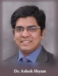
Vol 1 | Issue 1 |  December 2021 | page:01-02 | Dr. Ashok Shyam. DOI: 10.13107/jrs.2021.v01.i01.001


Author: Dr. Ashok Shyam [1,2]

[1] Department of Orthopaedic, Sancheti Institute for Orthopaedics and Rehabilitation, India.
[2] Chief Researcher, Orthopaedic Research Group, India.

Address of Correspondence
Dr. Ashok Shyam, MS Ortho.
Department of Orthopaedic, Sancheti Institute for Orthopaedics and Rehabilitation, Pune, Maharashtra, India.
Email: drashokshyam@gmail.com


Editorial:

Journal of Regenerative Science: A Step towards a more Biological Future

Biology defines and forms basis of every disease and treatment modality, it is the fundamental on which the entire science of medicine stands upon.

Restoration of Biology is essentially the core principle of Regenerative medicine. Regenerative Medicine is defined as “Regenerative Medicine is an emerging interdisciplinary field of research and clinical applications focused on the repair, replacement or regeneration of cells, tissues or organs to restore impaired function resulting from any cause, including congenital defects, disease, trauma and aging.” [1]. From the definition is clear the optimal restoration of functional biology is the aim of Regenerative medicine as opposed to clinical medicine where most of the focus lies on treating the symptoms or the external causative factors.

Regenerative medicine is a multidisciplinary branch which includes principles from engineering as well as biological sciences to utilise the innate ability of body to heal itself. A major part of this branch is dedicated to harvesting this ability while other parts are focussed on enhancing this ability or even replacing this ability by artificial means. The fact that earliest use of regenerative medicine was in early 1900 to prolong life or more specifically to slow the aging process. Although the term Regenerative medicine was coined in 1992 it was made popular only in 1999 and has seen the attention as well as applications in the field grow exponentially since then [2].

The scope of regenerative medicine is ever expanding but broadly focused on tissue engineering and tissue regeneration [3]. The Journal of Regenerative Science will focus more on tissue regeneration part, although advances and research in tissue engineering will receive special attention. When talks about this journal started few months back with Dr Daniel Moya, we were talking mostly in terms of tissue regeneration specifically non-invasive tissue regeneration using shockwaves. We had together conducted special webinars and scheduled talk on Shockwave therapies on our portal OrthoTV where we go incredible response to the concept. Feedback from viewers and especially orthopaedic surgeons was fantastic. We realised that this specific branch of regenerative medicine can be utilised quite effectively in musculoskeletal areas and would create a lot of positive impact on patient outcomes too. From this realisation stemmed the idea of creating a dedicated academic platform in form of Journal of Regenerative Science. It took us time to solidify the idea and find people who are as passionate as us about the subject. It is our great privilege to introduce the Editorial board of the Journal with the launch of first issue of JRS. We planned the first issue in two months and thanks to great effort by every author, we were able to bring out the first issue is a very short time. The issue is an excellent collection of invited articles form experts from across the globe and we are sure that reader will find value in reading these articles.

Regenerative science is a very futuristic branch, and it is still in its development phase. This is the stage where both the branch and the practitioner of the branch need to support each other through good research. For any new branch, current research is the foundation on which the future will be decided on. JRS is aimed to provide a peer reviewed platform where researchers can publish their research and help in growth of the specialty. JRS editorial board and reviewers list consists of great experts in the field and more will be joining us. This will help in maintaining highest quality of articles and publication in the Journal. At the same time, we wish to keep the focus of the articles to practical patient care too. Keeping both the academic as well as clinical aspect in mind we would like to extend an open invitation to all researchers in the field of Regenerative science to submit their work to the Journal.

The first issue is in your hands, and it is a work of lot of Love and hard work form authors and the editorial team. We sincerely hope that you will enjoy reading the articles and send us your feedback.

Wishing you a Very Safe and Happy New Year.


Dr. Ashok Shyam, MS Ortho


How to Cite this article: Shyam A. Journal of Regenerative Science: A Step towards a more Biological Future.  Journal of Regenerative Science | December 2021; 1(1):01-02.


  Dr. Ashok Shyam, MS Ortho

(Abstract    Full Text HTML)   (Download PDF)


Why a New Journal on Regenerative Medicine?

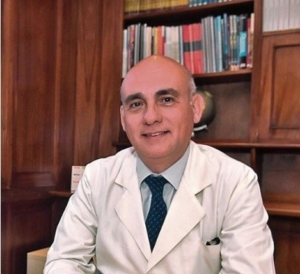
Vol 1 | Issue 1 |  December 2021 | page:03 | Dr. Daniel Moya. DOI: 10.13107/jrs.2021.v01.i01.003


Author: Dr. Daniel Moya [1]

[1] Department of Orthopaedic, Servicio de Ortopedia y Traumatología, Hospital Británico de Buenos Aires, Buenos Aires, Argentina.

Address of Correspondence
Dr. Daniel Moya, MD,.
Department of Orthopaedic, Servicio de Ortopedia y Traumatología, Hospital Británico de Buenos Aires, Buenos Aires, Argentina..
Email: drdanielmoya@yahoo.com.ar


Editorial:

Why a New Journal on Regenerative Medicine?

“Strange times are these in which we live, when old and young are taught falsehoods in school. And the person that dares to tell the truth is called at once a lunatic and fool.” It sounds current, however, it was stated by Plato almost 2400 years ago.

Obviously, the collision between old and new ideas, different points of view, and scientific approaches has always been conflictive. In our times this situation is exacerbated by the enormous generation of information and by the indiscriminate access to it.

Educational offer includes scientific societies, universities, predatory editors, industry, and even beginners, who simply by having access to social media become opinion makers. The tenuous limits between scientific information, marketing, and entertainment have their maximum expression in the field of social media.

What can we do? The answer also comes from the golden age of Greece: The scientific method. Its basis is the search for truth. The truth cannot be modified by biases.

The findings of observation and experimentation should be disseminated.

Despite isolated initiatives, there has not been a coordinated effort or an editorial forum dedicated to techniques such as the use of mechanical waves in non-invasive regenerative medicine. That is the reason of this publication. This Journal will also include information on minimally invasive, and invasive techniques as long as they are related to tissue regeneration.

Any applied treatment has no value if it is not based on a precise diagnosis, so it will also be of interest to this Journal to include not only therapeutic concepts but also the discussion of the diagnostic criteria and the most commonly used complementary tests.

The heart and soul of a publication are the authors. I will be eternally grateful to them for trusting in this project and enthusiastically joining this first issue.

The circuit will be completed with readers, their opinion and contribution will be valued. We will develop a section dedicated to this. Interaction with the readers will surely give the publication more dynamics and enrich it.

It would have been impossible to make this initiative a reality without the encouragement and trust of Dr. Ashok Shyham Pasi, the technical and scientific contribution of The Indian Orthopedic Research Group, the support of the Ibero Latin American Federation of Shockwaves and Tissue Engineering, and the editing input of Ms. Kajal Mandalia.

If we manage to improve the practice of a colleague or benefit the quality of life of a patient anywhere in the world, our mission will be fulfilled.


Dr. Daniel Moya, MD


How to Cite this article: Moya D. Why a New Journal on Regenerative Medicine?.  Journal of Regenerative Science | December 2021; 1(1):3.


  Dr. Daniel Moya, MD

(Abstract    Full Text HTML)   (Download PDF)


Blog Post Title

What goes into a blog post? Helpful, industry-specific content that: 1) gives readers a useful takeaway, and 2) shows you’re an industry expert.

Use your company’s blog posts to opine on current industry topics, humanize your company, and show how your products and services can help people.